个人简介
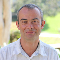
Pascal Egea received is B.S. in Molecular and Cellular Biology from the Ecole Normale Supérieure de Lyon (Lyon, France) in 1991. He then went to graduate school at the Université Louis Pasteur (Strasbourg, France) where he studied the structure and function of the retinoic acid receptors in the laboratory of Dr Dino Moras. After completing is Ph.D. in 1999, Pascal went to the University of California in San Francisco for post-doctoral training under the co-mentorship of Professors Robert Stroud and Peter Walter; during this period he studied the signal recognition particle and protein translocation pathways. Pascal joined the Department of Biological Chemistry as an assistant professor in the fall of 2009.
研究领域
 查看导师新发文章
(温馨提示:请注意重名现象,建议点开原文通过作者单位确认)
Structure and Function of Eukaryotic Membrane Protein Complexes Linked To Diseases
Most biological functions come about through the orchestrated interactions between macromolecules assembled into complexes. Membrane proteins account for ~25% of any given proteome and are the targets for ~50% of the therapeutic drugs in use today; yet very few three-dimensional structures of these valuable targets are known. Research in the Egea lab focuses on two specific eukaryotic membrane protein complexes studied at the level of their molecular structure to elucidate their mechanism of action and cellular functions. Because of their extreme complexity (in terms of molecular size and conformational flexibility) we apply a hybrid methods strategy to tackle these challenging assemblies. Our computational and structural approach combines X-ray crystallography to cryo-electron microscopy and solution scattering to close the resolution gap existing between the crystal structures of individual subunits and the experimental reconstructions of larger complexes and further validate our models.
Targeting a protein export complex for drug design in Plasmodium falciparum, the parasite causing malaria. Malaria is the world’s most important disease caused by a parasite. Nearly 10% of the world’s population is infected with malaria, which kills more than one million persons each year, with vast social and economic consequences. Modern research on malaria seeks to identify biological processes that are unique to the parasite. Such processes once characterized can be targeted for drug design. Pathogenesis in malaria almost entirely occurs at the red blood cell stage. The ability of the parasite to infect, remodel, and replicate in human erythrocytes is central to the pathogenesis of the disease. These processes are mediated in part by a large number of secreted essential parasite proteins that are transported across the vacuolar membrane via a parasite-derived translocon and delivered into the erythrocyte cytoplasm. The Plasmodium Translocon of Exported Proteins (PTEX) is a newly discovered membrane protein complex solely responsible for this export and thus represents an Achilles heel in the parasitic life cycle. This pathway is specific to the Plasmodium. Because vacuolar protein export is essential in Plasmodium, this process represents an ideal target for the design of novel therapies to treat and ultimately contribute to the eradication of the disease. The challenges posed by malaria reside in the rapid and recurrent emergence of resistance to existing anti-malarial therapies and the overall poor chemical diversity of the drugs at hand. PTEX is a large (2.5 MDa) membrane-bound complex composed of at least five subunits. We seek to characterize the structure of some of its key components to understand the mechanisms of export and guide the rational-design of novel classes of anti-malarial drugs: specific inhibitors of parasitic protein export.
The structure and functions of an inter-organelle tethering complex. How and why organelles communicate within the eukaryotic cell? Eukaryotic cells are characterized by a complex and interconnected network of membrane-bound organelles. Although cellular compartmentalization enables the efficient segregation of metabolic processes, the membranes that delineate these organelles impose a physical barrier impeding the exchange of matter and information. To overcome this problem, eukaryotic cells exploit membrane contact sites (MCSs), or regions of proximity between two organelles, to allow the transfer of metabolites. These MCSs serve as nexuses for the exchange of lipids and small molecules crucial to cellular life and homeostasis. Various protein complexes, such as the endoplasmic reticulum-mitochondrial encounter structure (ERMES), function as dynamic molecular tethers between organelles. ERMES is composed of five subunits; the absence of structural data has precluded answering questions such as how the subunits are organized to function as a tether, and whether they do indeed bind and exchange lipids at MCSs. Ultimately, the lack of structural insight undermines the understanding of function. We thus seek to reconstitute the entire ERMES. Although we use yeast as a model organism, MCSs are present in all eukaryotic cells. In humans, dysfunctional MCSs are associated with cancer and neurodegenerative diseases such as Parkinson and Alzheimer. Thus, answering a basic science question by studying a model system from yeast will have profound implications in human health sciences.


 京公网安备 11010802027423号