当前位置:
X-MOL 学术
›
ACS Appl. Mater. Interfaces
›
论文详情
Our official English website, www.x-mol.net, welcomes your
feedback! (Note: you will need to create a separate account there.)
Unraveling the Morphology–Function Relationships of Polyamide Membranes Using Quantitative Electron Tomography
ACS Applied Materials & Interfaces ( IF 8.3 ) Pub Date : 2019-01-24 00:00:00 , DOI: 10.1021/acsami.8b20826
Xiaohui Song , John W. Smith , Juyeong Kim , Nestor J. Zaluzec 1 , Wenxiang Chen , Hyosung An , Jordan M. Dennison , David G. Cahill , Matthew A. Kulzick 2 , Qian Chen
ACS Applied Materials & Interfaces ( IF 8.3 ) Pub Date : 2019-01-24 00:00:00 , DOI: 10.1021/acsami.8b20826
Xiaohui Song , John W. Smith , Juyeong Kim , Nestor J. Zaluzec 1 , Wenxiang Chen , Hyosung An , Jordan M. Dennison , David G. Cahill , Matthew A. Kulzick 2 , Qian Chen
Affiliation
|
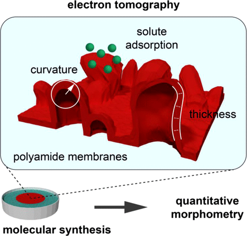
An understanding of how complex nanoscale morphologies emerge from synthesis would offer powerful strategies to construct soft materials with designed structures and functions. However, these kinds of morphologies have proven difficult to characterize, and therefore manipulate, because they are three-dimensional (3D), nanoscopic, and often highly irregular. Here, we studied polyamide (PA) membranes used in wastewater reclamation as a prime example of this challenge. Using electron tomography and quantitative morphometry, we reconstructed the nanoscale morphology of 3D crumples and voids in PA membranes for the first time. Various parameters governing film transport properties, such as surface-to-volume ratio and mass-per-area, were measured directly from the reconstructed membrane structure. In addition, we extracted information inaccessible by other means. For example, 3D reconstruction shows that membrane nanostructures are formed from PA layers 15–20 nm thick folding into 3D crumples which envelope up to 30% void by volume. Mapping local curvature and thickness in 3D quantitatively groups these crumples into three classes, “domes”, “dimples”, and “clusters”, each being a distinct type of microenvironment. Elemental mapping of metal ion adsorption across the film demonstrates that these previously missed parameters are relevant to membrane performance. This imaging–morphometry platform can be applicable to other nanoscale soft materials and potentially suggests engineering strategies based directly on synthesis–morphology–function relationships.
中文翻译:

使用定量电子层析成像技术揭示聚酰胺膜的形态-功能关系
对合成过程中如何产生复杂的纳米级形态的理解将为构造具有设计结构和功能的软材料提供强有力的策略。但是,由于这些形态是三维(3D)的,纳米的且通常高度不规则的,因此已被证明难以表征,因此难以操作。在这里,我们研究了废水回收中使用的聚酰胺(PA)膜作为这一挑战的主要例证。使用电子断层扫描和定量形态计量学,我们首次重建了PA膜中3D皱纹和空隙的纳米级形态。直接从重建的膜结构中测量了控制膜传输特性的各种参数,例如表面积与体积之比和单位面积质量。此外,我们提取了其他方式无法访问的信息。例如,3D重建表明,膜纳米结构是由15-20 nm厚的PA层折叠成3D皱褶形成的,这些皱褶包裹了30%的体积空隙。在3D模式下绘制局部曲率和厚度,将这些皱褶定量地分为三类,“圆顶”,“酒窝”和“簇”,每种都是微环境的不同类型。膜上金属离子吸附的元素图谱表明,这些先前遗漏的参数与膜性能有关。这种成像形态测量平台可适用于其他纳米级软材料,并可能直接基于合成,形态与功能之间的关系提出工程策略。3D重建表明,膜纳米结构是由15-20 nm厚的PA层折叠成3D皱褶形成的,这些皱褶包裹了30%的体积空隙。在3D模式下绘制局部曲率和厚度,将这些皱褶定量地分为三类,“圆顶”,“酒窝”和“簇”,每种都是微环境的不同类型。膜上金属离子吸附的元素图谱表明,这些先前遗漏的参数与膜性能有关。这种成像形态学平台可以适用于其他纳米级软材料,并可能直接基于合成,形态学,功能关系提出工程策略。3D重建表明,膜纳米结构是由15-20 nm厚的PA层折叠成3D皱褶形成的,这些皱褶包裹了30%的体积空隙。在3D模式下绘制局部曲率和厚度,将这些皱褶定量地分为三类,“圆顶”,“酒窝”和“簇”,每种都是微环境的不同类型。膜上金属离子吸附的元素图谱表明,这些先前遗漏的参数与膜性能有关。这种成像形态测量平台可适用于其他纳米级软材料,并可能直接基于合成,形态与功能之间的关系提出工程策略。在3D模式下绘制局部曲率和厚度,将这些皱褶定量地分为三类,“圆顶”,“酒窝”和“簇”,每种都是微环境的不同类型。膜上金属离子吸附的元素图谱表明,这些先前遗漏的参数与膜性能有关。这种成像形态测量平台可适用于其他纳米级软材料,并可能直接基于合成,形态与功能之间的关系提出工程策略。在3D模式下绘制局部曲率和厚度,将这些皱褶定量地分为三类,“圆顶”,“酒窝”和“簇”,每种都是微环境的不同类型。膜上金属离子吸附的元素图谱表明,这些先前遗漏的参数与膜性能有关。这种成像形态测量平台可适用于其他纳米级软材料,并可能直接基于合成,形态与功能之间的关系提出工程策略。
更新日期:2019-01-24
中文翻译:
使用定量电子层析成像技术揭示聚酰胺膜的形态-功能关系
对合成过程中如何产生复杂的纳米级形态的理解将为构造具有设计结构和功能的软材料提供强有力的策略。但是,由于这些形态是三维(3D)的,纳米的且通常高度不规则的,因此已被证明难以表征,因此难以操作。在这里,我们研究了废水回收中使用的聚酰胺(PA)膜作为这一挑战的主要例证。使用电子断层扫描和定量形态计量学,我们首次重建了PA膜中3D皱纹和空隙的纳米级形态。直接从重建的膜结构中测量了控制膜传输特性的各种参数,例如表面积与体积之比和单位面积质量。此外,我们提取了其他方式无法访问的信息。例如,3D重建表明,膜纳米结构是由15-20 nm厚的PA层折叠成3D皱褶形成的,这些皱褶包裹了30%的体积空隙。在3D模式下绘制局部曲率和厚度,将这些皱褶定量地分为三类,“圆顶”,“酒窝”和“簇”,每种都是微环境的不同类型。膜上金属离子吸附的元素图谱表明,这些先前遗漏的参数与膜性能有关。这种成像形态测量平台可适用于其他纳米级软材料,并可能直接基于合成,形态与功能之间的关系提出工程策略。3D重建表明,膜纳米结构是由15-20 nm厚的PA层折叠成3D皱褶形成的,这些皱褶包裹了30%的体积空隙。在3D模式下绘制局部曲率和厚度,将这些皱褶定量地分为三类,“圆顶”,“酒窝”和“簇”,每种都是微环境的不同类型。膜上金属离子吸附的元素图谱表明,这些先前遗漏的参数与膜性能有关。这种成像形态学平台可以适用于其他纳米级软材料,并可能直接基于合成,形态学,功能关系提出工程策略。3D重建表明,膜纳米结构是由15-20 nm厚的PA层折叠成3D皱褶形成的,这些皱褶包裹了30%的体积空隙。在3D模式下绘制局部曲率和厚度,将这些皱褶定量地分为三类,“圆顶”,“酒窝”和“簇”,每种都是微环境的不同类型。膜上金属离子吸附的元素图谱表明,这些先前遗漏的参数与膜性能有关。这种成像形态测量平台可适用于其他纳米级软材料,并可能直接基于合成,形态与功能之间的关系提出工程策略。在3D模式下绘制局部曲率和厚度,将这些皱褶定量地分为三类,“圆顶”,“酒窝”和“簇”,每种都是微环境的不同类型。膜上金属离子吸附的元素图谱表明,这些先前遗漏的参数与膜性能有关。这种成像形态测量平台可适用于其他纳米级软材料,并可能直接基于合成,形态与功能之间的关系提出工程策略。在3D模式下绘制局部曲率和厚度,将这些皱褶定量地分为三类,“圆顶”,“酒窝”和“簇”,每种都是微环境的不同类型。膜上金属离子吸附的元素图谱表明,这些先前遗漏的参数与膜性能有关。这种成像形态测量平台可适用于其他纳米级软材料,并可能直接基于合成,形态与功能之间的关系提出工程策略。


 京公网安备 11010802027423号