Our official English website, www.x-mol.net, welcomes your
feedback! (Note: you will need to create a separate account there.)
Biomimetic Dextran-Based Hydrogel Layers for Cell Micropatterning over Large Areas Using the FluidFM BOT Technology.
Langmuir ( IF 3.7 ) Pub Date : 2019-01-17 00:00:00 , DOI: 10.1021/acs.langmuir.8b03249 Andras Saftics 1 , Barbara Türk 1 , Attila Sulyok , Norbert Nagy , Tamás Gerecsei 2 , Inna Szekacs , Sándor Kurunczi , Robert Horvath
Langmuir ( IF 3.7 ) Pub Date : 2019-01-17 00:00:00 , DOI: 10.1021/acs.langmuir.8b03249 Andras Saftics 1 , Barbara Türk 1 , Attila Sulyok , Norbert Nagy , Tamás Gerecsei 2 , Inna Szekacs , Sándor Kurunczi , Robert Horvath
Affiliation

|
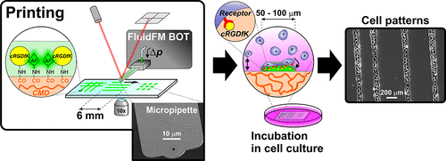
Micropatterning of living single cells and cell clusters over millimeter–centimeter scale areas is of high demand in the development of cell-based biosensors. Micropatterning methodologies require both a suitable biomimetic support and a printing technology. In this work, we present the micropatterning of living mammalian cells on carboxymethyl dextran (CMD) hydrogel layers using the FluidFM BOT technology. In contrast to the ultrathin (few nanometers thick in the dry state) CMD films generally used in label-free biosensor applications, we developed CMD layers with thicknesses of several tens of nanometers in order to provide support for the controlled adhesion of living cells. The fabrication method and detailed characterization of the CMD layers are also described. The antifouling ability of the CMD surfaces is demonstrated by in situ optical waveguide lightmode spectroscopy measurements using serum modeling proteins with different electrostatic properties and molecular weights. Cell micropatterning on the CMD surface was obtained by printing cell adhesion mediating cRGDfK peptide molecules (cyclo(Arg-Gly-Asp-d-Phe-Lys)) directly from aqueous solution using microchanneled cantilevers with subsequent incubation of the printed surfaces in the living cell culture. Uniquely, we present cell patterns with different geometries (spot, line, and grid arrays) covering both micrometer and millimeter–centimeter scale areas. The adhered patterns were analyzed by phase contrast microscopy and the adhesion process on the patterns was real-time monitored by digital holographic microscopy, enabling to quantify the survival and migration of cells on the printed cRGDfK arrays.
中文翻译:

基于仿生葡聚糖的水凝胶层,使用FluidFM BOT技术在大面积上进行细胞微图案化。
在基于细胞的生物传感器的开发中,对毫米级以上的单个活细胞和细胞簇的微图案化提出了很高的要求。微图案化方法学既需要合适的仿生载体,也需要印刷技术。在这项工作中,我们介绍了使用FluidFM BOT技术在羧甲基右旋糖酐(CMD)水凝胶层上对哺乳动物活细胞的微图案化。与通常用于无标签生物传感器应用的超薄(干燥状态下只有几纳米厚)的CMD膜相反,我们开发了厚度为几十纳米的CMD层,以便为可控的活细胞粘附提供支持。还描述了CMD层的制造方法和详细特性。通过使用具有不同静电特性和分子量的血清建模蛋白,通过原位光波导光模光谱测量法证明了CMD表面的防污能力。通过打印介导cRGDfK肽分子(环(Arg-Gly-Asp-d -Phe-Lys)),直接使用微通道悬臂从水溶液中提取,然后在活细胞培养物中孵育印刷的表面。独特的是,我们展示了具有不同几何形状(点,线和网格阵列)的单元模式,覆盖了微米和毫米厘米级的区域。通过相差显微镜分析粘附的图案,并通过数字全息显微镜实时监测图案上的粘附过程,从而能够量化打印的cRGDfK阵列上细胞的存活和迁移。
更新日期:2019-01-17
中文翻译:

基于仿生葡聚糖的水凝胶层,使用FluidFM BOT技术在大面积上进行细胞微图案化。
在基于细胞的生物传感器的开发中,对毫米级以上的单个活细胞和细胞簇的微图案化提出了很高的要求。微图案化方法学既需要合适的仿生载体,也需要印刷技术。在这项工作中,我们介绍了使用FluidFM BOT技术在羧甲基右旋糖酐(CMD)水凝胶层上对哺乳动物活细胞的微图案化。与通常用于无标签生物传感器应用的超薄(干燥状态下只有几纳米厚)的CMD膜相反,我们开发了厚度为几十纳米的CMD层,以便为可控的活细胞粘附提供支持。还描述了CMD层的制造方法和详细特性。通过使用具有不同静电特性和分子量的血清建模蛋白,通过原位光波导光模光谱测量法证明了CMD表面的防污能力。通过打印介导cRGDfK肽分子(环(Arg-Gly-Asp-d -Phe-Lys)),直接使用微通道悬臂从水溶液中提取,然后在活细胞培养物中孵育印刷的表面。独特的是,我们展示了具有不同几何形状(点,线和网格阵列)的单元模式,覆盖了微米和毫米厘米级的区域。通过相差显微镜分析粘附的图案,并通过数字全息显微镜实时监测图案上的粘附过程,从而能够量化打印的cRGDfK阵列上细胞的存活和迁移。


 京公网安备 11010802027423号