当前位置:
X-MOL 学术
›
ACS Appl. Mater. Interfaces
›
论文详情
Our official English website, www.x-mol.net, welcomes your
feedback! (Note: you will need to create a separate account there.)
Fibrin Nanoparticles Coupled with Keratinocyte Growth Factor Enhance the Dermal Wound-Healing Rate
ACS Applied Materials & Interfaces ( IF 8.3 ) Pub Date : 2019-01-03 00:00:00 , DOI: 10.1021/acsami.8b21056 Ismaeel Muhamed 1, 2 , Erin P. Sproul 1, 2 , Frances S. Ligler 1, 2 , Ashley C. Brown 1, 2
ACS Applied Materials & Interfaces ( IF 8.3 ) Pub Date : 2019-01-03 00:00:00 , DOI: 10.1021/acsami.8b21056 Ismaeel Muhamed 1, 2 , Erin P. Sproul 1, 2 , Frances S. Ligler 1, 2 , Ashley C. Brown 1, 2
Affiliation

|
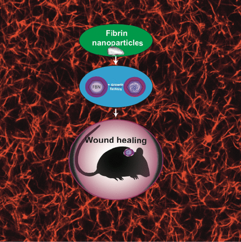
Expediting the wound-healing process is critical for patients chronically ill from nonhealing wounds and diseases such as hemophilia or diabetes or who have suffered trauma including easily infected open wounds. FDA-approved external tissue sealants include the topical application of fibrin gels, which can be 500 times denser than natural fibrin clots. With lower clot porosity and higher polymerization rates than physiologically formed fibrin clots, the commercial gels quickly stop blood loss but impede the later clot degradation kinetics and thus retard tissue-healing rates. The fibrin nanoparticles (FBNs) described here are constructed from physiologically relevant fibrin concentrations that support new tissue and dermal wound scaffold formation when coupled with growth factors. The FBNs, synthesized in a microfluidic droplet generator, support cell adhesion and traction generation, and when coupled to keratinocyte growth factor (KGF), support cell migration and in vivo wound healing. The FBN–KGF particles enhance cell migration in vitro greater than FBN alone or free KGF and also improve healing outcomes in a murine full thickness injury model compared to saline, bulk fibrin sealant, free KGF, or bulk fibrin mixed with KGF treatments. Furthermore, FBN can be potentially administered with other tissue-healing factors and inflammatory mediators to improve wound-healing outcomes.
中文翻译:

纤维蛋白纳米颗粒与角质形成细胞生长因子结合提高皮肤伤口愈合率
对于那些因伤口不愈合和血友病或糖尿病等疾病而长期患病或遭受包括容易感染的开放性伤口在内的创伤的患者而言,加快伤口的愈合过程至关重要。FDA批准的外部组织密封剂包括局部应用的血纤蛋白凝胶,其密度可比天然血纤蛋白凝块高500倍。与生理上形成的血纤蛋白凝块相比,凝块孔隙率较低,聚合速率较高,市售的凝胶可迅速停止失血,但会阻碍后来的凝块降解动力学,从而阻碍组织的愈合速度。本文所述的纤维蛋白纳米颗粒(FBN)由生理相关的纤维蛋白浓度构建而成,当与生长因子结合时,该浓度可支持新组织和皮肤伤口支架的形成。在微流液滴发生器中合成的FBN,支持细胞粘附和牵引力生成,并与角质形成细胞生长因子(KGF)结合时,支持细胞迁移和体内伤口愈合。与盐水,散装纤维蛋白封闭剂,游离KGF或散装纤维蛋白结合KGF治疗相比,FBN–KGF颗粒在体外的细胞迁移比单独的FBN或游离KGF更大,并且还改善了鼠的全层损伤模型的愈合结果。此外,FBN可以与其他组织愈合因子和炎性介质一起使用,以改善伤口愈合效果。与盐水,块状纤维蛋白封闭剂,游离KGF或块状纤维蛋白与KGF处理混合相比,FBN–KGF颗粒在体外的细胞迁移比单独的FBN或游离KGF更大,并且还改善了鼠的全层损伤模型的愈合结果。此外,FBN可以与其他组织愈合因子和炎性介质一起使用,以改善伤口愈合效果。与盐水,散装纤维蛋白封闭剂,游离KGF或散装纤维蛋白结合KGF治疗相比,FBN–KGF颗粒在体外的细胞迁移比单独的FBN或游离KGF更大,并且还改善了鼠的全层损伤模型的愈合结果。此外,FBN可以与其他组织愈合因子和炎性介质一起使用,以改善伤口愈合效果。
更新日期:2019-01-03
中文翻译:

纤维蛋白纳米颗粒与角质形成细胞生长因子结合提高皮肤伤口愈合率
对于那些因伤口不愈合和血友病或糖尿病等疾病而长期患病或遭受包括容易感染的开放性伤口在内的创伤的患者而言,加快伤口的愈合过程至关重要。FDA批准的外部组织密封剂包括局部应用的血纤蛋白凝胶,其密度可比天然血纤蛋白凝块高500倍。与生理上形成的血纤蛋白凝块相比,凝块孔隙率较低,聚合速率较高,市售的凝胶可迅速停止失血,但会阻碍后来的凝块降解动力学,从而阻碍组织的愈合速度。本文所述的纤维蛋白纳米颗粒(FBN)由生理相关的纤维蛋白浓度构建而成,当与生长因子结合时,该浓度可支持新组织和皮肤伤口支架的形成。在微流液滴发生器中合成的FBN,支持细胞粘附和牵引力生成,并与角质形成细胞生长因子(KGF)结合时,支持细胞迁移和体内伤口愈合。与盐水,散装纤维蛋白封闭剂,游离KGF或散装纤维蛋白结合KGF治疗相比,FBN–KGF颗粒在体外的细胞迁移比单独的FBN或游离KGF更大,并且还改善了鼠的全层损伤模型的愈合结果。此外,FBN可以与其他组织愈合因子和炎性介质一起使用,以改善伤口愈合效果。与盐水,块状纤维蛋白封闭剂,游离KGF或块状纤维蛋白与KGF处理混合相比,FBN–KGF颗粒在体外的细胞迁移比单独的FBN或游离KGF更大,并且还改善了鼠的全层损伤模型的愈合结果。此外,FBN可以与其他组织愈合因子和炎性介质一起使用,以改善伤口愈合效果。与盐水,散装纤维蛋白封闭剂,游离KGF或散装纤维蛋白结合KGF治疗相比,FBN–KGF颗粒在体外的细胞迁移比单独的FBN或游离KGF更大,并且还改善了鼠的全层损伤模型的愈合结果。此外,FBN可以与其他组织愈合因子和炎性介质一起使用,以改善伤口愈合效果。


 京公网安备 11010802027423号