当前位置:
X-MOL 学术
›
Eur. J. Clin. Nutr.
›
论文详情
Our official English website, www.x-mol.net, welcomes your
feedback! (Note: you will need to create a separate account there.)
CKD autophagy activation and skeletal muscle atrophy-a preliminary study of mitophagy and inflammation.
European Journal of Clinical Nutrition ( IF 3.6 ) Pub Date : 2019-01-03 , DOI: 10.1038/s41430-018-0381-x Yue Yue Zhang 1 , Li Jie Gu 1 , Juan Huang 1 , Min Chao Cai 1 , Hong Lei Yu 1 , Wei Zhang 1 , Jin Fang Bao 1 , Wei Jie Yuan 1
European Journal of Clinical Nutrition ( IF 3.6 ) Pub Date : 2019-01-03 , DOI: 10.1038/s41430-018-0381-x Yue Yue Zhang 1 , Li Jie Gu 1 , Juan Huang 1 , Min Chao Cai 1 , Hong Lei Yu 1 , Wei Zhang 1 , Jin Fang Bao 1 , Wei Jie Yuan 1
Affiliation

|
BACKGROUND/OBJECTIVES
Long-lived proteins and organelles, such as mitochondria and the sarcoplasmic reticulum, are degraded by autophagy. However, the specific role of autophagy in chronic kidney disease (CKD) muscle atrophy is still undefined.
SUBJECTS/METHODS
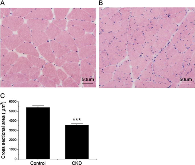
This was a cross-sectional study with 20 subjects and 11 controls. Autophagy induction was studied in human skeletal muscle biopsies from CKD patients and controls by comparing the cross-sectional areas of muscle fibers, protein, and mRNA expression of autophagy-related genes and the appearance of autophagosomes.
RESULTS
The cross-sectional area of muscle fibers was decreased in CKD patients as compared with the control group. CKD was associated with activated autophagy and mitophagy, as measured by the elevated mRNA and protein expression of BNIP3, (microtubule-associated proteins 1 A/1B light chain 3, also MAP1LC3) LC3, p62, PINK1, and PARKIN in the skeletal muscle and isolated mitochondria of the CKD group. Electron microscopy and immunohistofluorescence analysis showed mitochondrial engulfment by autophagosomes. Mitophagy was further demonstrated by the colocalization of LC3 and p62 puncta with the mitochondrial outer membrane protein TOM20. In addition, degradative FOXO3 (Forkhead box O3) was activated and synthetic mTOR (mammalian target of rapamycin) was inhibited, whereas the upstream mediators VPS34 (class III PI3-kinase) and AKT (protein kinase B, PKB) were activated in CKD patients.
CONCLUSIONS
Hyperactive autophagy and mitophagy may play important roles in CKD muscle atrophy. Autophagy was activated by FOXO3 translational factors in the skeletal muscle tissues of CKD patients, which maybe a new way of intervention for CKD muscle atrophy.
中文翻译:

CKD自噬激活和骨骼肌萎缩——线粒体自噬和炎症的初步研究。
背景/目的 长寿的蛋白质和细胞器,例如线粒体和肌质网,会被自噬降解。然而,自噬在慢性肾脏病 (CKD) 肌肉萎缩中的具体作用仍未确定。受试者/方法 这是一项包含 20 名受试者和 11 名对照的横断面研究。通过比较自噬相关基因的肌纤维横截面积、蛋白质和 mRNA 表达以及自噬体的外观,在来自 CKD 患者和对照的人体骨骼肌活检中研究自噬诱导。结果CKD患者肌纤维横截面积较对照组减少。CKD 与活化的自噬和线粒体自噬相关,通过 BNIP3 的 mRNA 和蛋白表达升高来衡量,(微管相关蛋白 1 A/1B 轻链 3,还有 MAP1LC3)CKD 组骨骼肌和分离线粒体中的 LC3、p62、PINK1 和 PARKIN。电子显微镜和免疫组织荧光分析显示线粒体被自噬体吞噬。LC3 和 p62 puncta 与线粒体外膜蛋白 TOM20 的共定位进一步证明了线粒体自噬。此外,降解的 FOXO3(叉头盒 O3)被激活,合成的 mTOR(雷帕霉素的哺乳动物靶点)被抑制,而上游介质 VPS34(III 类 PI3 激酶)和 AKT(蛋白激酶 B,PKB)在 CKD 患者中被激活. 结论过度活跃的自噬和线粒体自噬可能在CKD肌肉萎缩中起重要作用。自噬被 CKD 患者骨骼肌组织中的 FOXO3 翻译因子激活,
更新日期:2019-01-26
中文翻译:

CKD自噬激活和骨骼肌萎缩——线粒体自噬和炎症的初步研究。
背景/目的 长寿的蛋白质和细胞器,例如线粒体和肌质网,会被自噬降解。然而,自噬在慢性肾脏病 (CKD) 肌肉萎缩中的具体作用仍未确定。受试者/方法 这是一项包含 20 名受试者和 11 名对照的横断面研究。通过比较自噬相关基因的肌纤维横截面积、蛋白质和 mRNA 表达以及自噬体的外观,在来自 CKD 患者和对照的人体骨骼肌活检中研究自噬诱导。结果CKD患者肌纤维横截面积较对照组减少。CKD 与活化的自噬和线粒体自噬相关,通过 BNIP3 的 mRNA 和蛋白表达升高来衡量,(微管相关蛋白 1 A/1B 轻链 3,还有 MAP1LC3)CKD 组骨骼肌和分离线粒体中的 LC3、p62、PINK1 和 PARKIN。电子显微镜和免疫组织荧光分析显示线粒体被自噬体吞噬。LC3 和 p62 puncta 与线粒体外膜蛋白 TOM20 的共定位进一步证明了线粒体自噬。此外,降解的 FOXO3(叉头盒 O3)被激活,合成的 mTOR(雷帕霉素的哺乳动物靶点)被抑制,而上游介质 VPS34(III 类 PI3 激酶)和 AKT(蛋白激酶 B,PKB)在 CKD 患者中被激活. 结论过度活跃的自噬和线粒体自噬可能在CKD肌肉萎缩中起重要作用。自噬被 CKD 患者骨骼肌组织中的 FOXO3 翻译因子激活,


 京公网安备 11010802027423号