当前位置:
X-MOL 学术
›
Nat. Plants
›
论文详情
Our official English website, www.x-mol.net, welcomes your
feedback! (Note: you will need to create a separate account there.)
A whole-cell electron tomography model of vacuole biogenesis in Arabidopsis root cells.
Nature Plants ( IF 15.8 ) Pub Date : 2019-Jan-01 , DOI: 10.1038/s41477-018-0328-1 Yong Cui 1 , Wenhan Cao 1 , Yilin He 1 , Qiong Zhao 1 , Mayumi Wakazaki 2 , Xiaohong Zhuang 1 , Jiayang Gao 1 , Yonglun Zeng 1 , Caiji Gao 1, 3 , Yu Ding 1, 4 , Hiu Yan Wong 1 , Wing Shing Wong 1 , Ham Karen Lam 1 , Pengfei Wang 1 , Takashi Ueda 5 , Marcela Rojas-Pierce 6 , Kiminori Toyooka 2 , Byung-Ho Kang 1 , Liwen Jiang 1, 7
Nature Plants ( IF 15.8 ) Pub Date : 2019-Jan-01 , DOI: 10.1038/s41477-018-0328-1 Yong Cui 1 , Wenhan Cao 1 , Yilin He 1 , Qiong Zhao 1 , Mayumi Wakazaki 2 , Xiaohong Zhuang 1 , Jiayang Gao 1 , Yonglun Zeng 1 , Caiji Gao 1, 3 , Yu Ding 1, 4 , Hiu Yan Wong 1 , Wing Shing Wong 1 , Ham Karen Lam 1 , Pengfei Wang 1 , Takashi Ueda 5 , Marcela Rojas-Pierce 6 , Kiminori Toyooka 2 , Byung-Ho Kang 1 , Liwen Jiang 1, 7
Affiliation
|
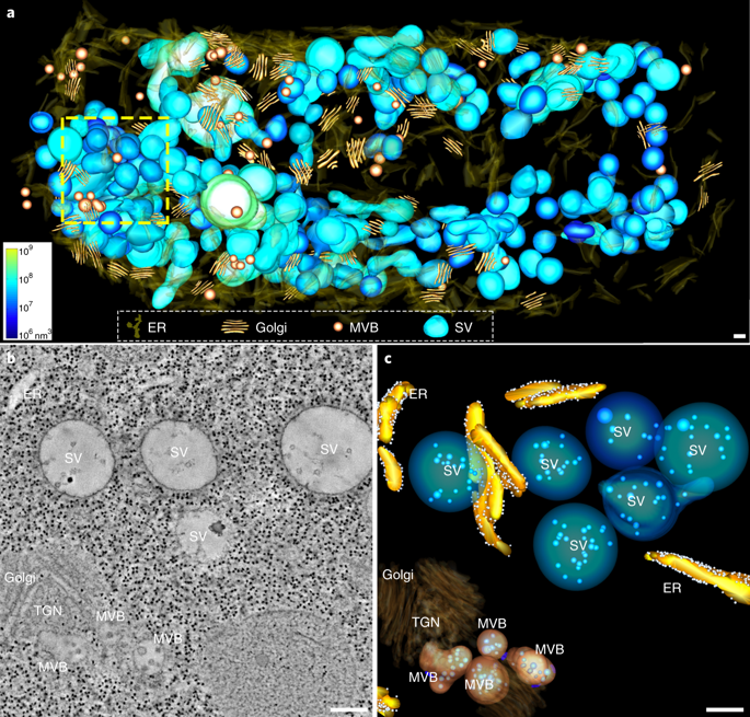
Plant vacuoles are dynamic organelles that play essential roles in regulating growth and development. Two distinct models of vacuole biogenesis have been proposed: separate vacuoles are formed by the fusion of endosomes, or the single interconnected vacuole is derived from the endoplasmic reticulum. These two models are based on studies of two-dimensional (2D) transmission electron microscopy and 3D confocal imaging, respectively. Here, we performed 3D electron tomography at nanometre resolution to illustrate vacuole biogenesis in Arabidopsis root cells. The whole-cell electron tomography analysis first identified unique small vacuoles (SVs; 400-1,000 nm in diameter) as nascent vacuoles in early developmental cortical cells. These SVs contained intraluminal vesicles and were mainly derived/matured from multivesicular body (MVB) fusion. The whole-cell vacuole models and statistical analysis on wild-type root cells of different vacuole developmental stages demonstrated that central vacuoles were derived from MVB-to-SV transition and subsequent fusions of SVs. Further electron tomography analysis on mutants defective in MVB formation/maturation or vacuole fusion demonstrated that central vacuole formation required functional MVBs and membrane fusion machineries.
中文翻译:

拟南芥根细胞液泡生物发生的全细胞电子断层扫描模型。
植物液泡是动态的细胞器,在调节生长和发育中起重要作用。已经提出了两种不同的液泡生物发生模型:单独的液泡由内体融合形成,或者单个相互连接的液泡来源于内质网。这两个模型分别基于二维 (2D) 透射电子显微镜和 3D 共焦成像的研究。在这里,我们以纳米分辨率进行了 3D 电子断层扫描,以说明拟南芥根细胞中的液泡生物发生。全细胞电子断层扫描分析首先将独特的小液泡(SV;直径 400-1,000 nm)鉴定为早期发育皮层细胞中的新生液泡。这些 SV 含有腔内囊泡,主要来源于多泡体 (MVB) 融合。对不同液泡发育阶段的野生型根细胞的全细胞液泡模型和统计分析表明,中央液泡来源于MVB-to-SV过渡和随后的SV融合。对 MVB 形成/成熟或液泡融合缺陷突变体的进一步电子断层扫描分析表明,中央液泡形成需要功能性 MVB 和膜融合机制。
更新日期:2019-01-26
中文翻译:
拟南芥根细胞液泡生物发生的全细胞电子断层扫描模型。
植物液泡是动态的细胞器,在调节生长和发育中起重要作用。已经提出了两种不同的液泡生物发生模型:单独的液泡由内体融合形成,或者单个相互连接的液泡来源于内质网。这两个模型分别基于二维 (2D) 透射电子显微镜和 3D 共焦成像的研究。在这里,我们以纳米分辨率进行了 3D 电子断层扫描,以说明拟南芥根细胞中的液泡生物发生。全细胞电子断层扫描分析首先将独特的小液泡(SV;直径 400-1,000 nm)鉴定为早期发育皮层细胞中的新生液泡。这些 SV 含有腔内囊泡,主要来源于多泡体 (MVB) 融合。对不同液泡发育阶段的野生型根细胞的全细胞液泡模型和统计分析表明,中央液泡来源于MVB-to-SV过渡和随后的SV融合。对 MVB 形成/成熟或液泡融合缺陷突变体的进一步电子断层扫描分析表明,中央液泡形成需要功能性 MVB 和膜融合机制。


 京公网安备 11010802027423号