Our official English website, www.x-mol.net, welcomes your
feedback! (Note: you will need to create a separate account there.)
Ultrasonography for lymph nodes metastasis identification in bitches with mammary neoplasms.
Scientific Reports ( IF 3.8 ) Pub Date : 2018-Dec-07 , DOI: 10.1038/s41598-018-34806-9 Priscila Silva , Ricardo Andres Ramirez Uscategui , Marjury Cristina Maronezi , Beatriz Gasser , Letícia Pavan , Igor Renan Honorato Gatto , Vivian Tavares de Almeida , Wilter Ricardo Russiano Vicente , Marcus Antônio Rossi Feliciano
Scientific Reports ( IF 3.8 ) Pub Date : 2018-Dec-07 , DOI: 10.1038/s41598-018-34806-9 Priscila Silva , Ricardo Andres Ramirez Uscategui , Marjury Cristina Maronezi , Beatriz Gasser , Letícia Pavan , Igor Renan Honorato Gatto , Vivian Tavares de Almeida , Wilter Ricardo Russiano Vicente , Marcus Antônio Rossi Feliciano

|
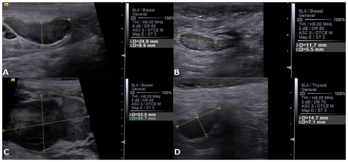
The aim of this study was to evaluate and compare the diagnostic accuracy of B-mode, Doppler ultrasonography and Acoustic Radiation Force Impulse (ARFI) elastography in the identification of axillary and inguinal lymph nodes metastasis in bitches with mammary neoplasms. The axillary (n = 96) and inguinal (n = 100) lymph nodes of 100 bitches were evaluated using B-Mode, Colour Doppler and ARFI-elastography. After this evaluation, mastectomy and lymph nodes excision were performed and these structures were histologically classified as free, reactive or metastatic. Ultrasonographic parameters were compared by Chi-Square or ANOVA tests and if they are significant, discriminative power analysis according to histopathological classification was performed (ROC analysis). The ARFI-elastography shear wave velocity (SWV) enabled metastasis identification in inguinal (sensitivity 95% specificity 87%) and axillary lymph nodes (sensitivity 100% specificity 94%). While B-Mode ultrasound Short/Long axis ratio evaluation of inguinal and axillary lymph nodes only resulted in a sensitivity around of 71% and specificity of 55%. In conclusion, B-Mode ultrasonography may contribute to diagnosis of metastasis in axillary and inguinal lymph nodes of bitches affected by mammary neoplasm with limited accuracy, while SWV evaluation proved to be an excellent diagnosis tool, which allows differentiation between free, reactive and tumour metastatic lymph nodes.
中文翻译:

超声检查在乳腺肿瘤母犬中的淋巴结转移识别。
这项研究的目的是评估和比较B型,多普勒超声检查和声辐射力脉冲(ARFI)弹性成像在乳腺肿瘤母犬腋窝和腹股沟淋巴结转移中的诊断准确性。使用B型,彩色多普勒和ARFI弹性成像技术评估了100个母犬的腋窝淋巴结(n = 96)和腹股沟淋巴结(n = 100)。评估后,进行乳房切除术和淋巴结切除术,这些结构在组织学上分类为游离,反应性或转移性。通过卡方检验或方差分析检验超声参数,如果它们显着,则根据组织病理学分类进行鉴别力分析(ROC分析)。ARFI弹力图剪切波速度(SWV)能够识别腹股沟(敏感性95%特异性87%)和腋窝淋巴结(敏感性100%特异性94%)中的转移。B型超声对腹股沟和腋窝淋巴结的短/长轴比率的评估仅导致大约71%的敏感性和55%的特异性。总之,B型超声检查可能有助于诊断受乳腺肿瘤影响的母犬腋窝和腹股沟淋巴结转移,但准确性有限,而SWV评估是一种出色的诊断工具,可以区分游离,反应性和肿瘤转移淋巴结。B型超声对腹股沟和腋窝淋巴结的短/长轴比率的评估仅导致大约71%的敏感性和55%的特异性。总之,B型超声检查可能有助于诊断受乳腺肿瘤影响的母犬腋窝和腹股沟淋巴结转移,但准确性有限,而SWV评估是一种出色的诊断工具,可以区分游离,反应性和肿瘤转移淋巴结。B型超声对腹股沟和腋窝淋巴结的短/长轴比率的评估仅导致大约71%的敏感性和55%的特异性。总之,B型超声检查可能有助于诊断受乳腺肿瘤影响的母犬腋窝和腹股沟淋巴结转移,但准确性有限,而SWV评估是一种出色的诊断工具,可以区分游离,反应性和肿瘤转移淋巴结。
更新日期:2018-12-07
中文翻译:

超声检查在乳腺肿瘤母犬中的淋巴结转移识别。
这项研究的目的是评估和比较B型,多普勒超声检查和声辐射力脉冲(ARFI)弹性成像在乳腺肿瘤母犬腋窝和腹股沟淋巴结转移中的诊断准确性。使用B型,彩色多普勒和ARFI弹性成像技术评估了100个母犬的腋窝淋巴结(n = 96)和腹股沟淋巴结(n = 100)。评估后,进行乳房切除术和淋巴结切除术,这些结构在组织学上分类为游离,反应性或转移性。通过卡方检验或方差分析检验超声参数,如果它们显着,则根据组织病理学分类进行鉴别力分析(ROC分析)。ARFI弹力图剪切波速度(SWV)能够识别腹股沟(敏感性95%特异性87%)和腋窝淋巴结(敏感性100%特异性94%)中的转移。B型超声对腹股沟和腋窝淋巴结的短/长轴比率的评估仅导致大约71%的敏感性和55%的特异性。总之,B型超声检查可能有助于诊断受乳腺肿瘤影响的母犬腋窝和腹股沟淋巴结转移,但准确性有限,而SWV评估是一种出色的诊断工具,可以区分游离,反应性和肿瘤转移淋巴结。B型超声对腹股沟和腋窝淋巴结的短/长轴比率的评估仅导致大约71%的敏感性和55%的特异性。总之,B型超声检查可能有助于诊断受乳腺肿瘤影响的母犬腋窝和腹股沟淋巴结转移,但准确性有限,而SWV评估是一种出色的诊断工具,可以区分游离,反应性和肿瘤转移淋巴结。B型超声对腹股沟和腋窝淋巴结的短/长轴比率的评估仅导致大约71%的敏感性和55%的特异性。总之,B型超声检查可能有助于诊断受乳腺肿瘤影响的母犬腋窝和腹股沟淋巴结转移,但准确性有限,而SWV评估是一种出色的诊断工具,可以区分游离,反应性和肿瘤转移淋巴结。


 京公网安备 11010802027423号