Our official English website, www.x-mol.net, welcomes your
feedback! (Note: you will need to create a separate account there.)
Pulsed interleaved excitation-based line-scanning spatial correlation spectroscopy (PIE-lsSCS).
Scientific Reports ( IF 3.8 ) Pub Date : 2018-Nov-13 , DOI: 10.1038/s41598-018-35146-4 Xiang Gao 1 , Peng Gao 1, 2 , Benedikt Prunsche 1 , Karin Nienhaus 1 , Gerd Ulrich Nienhaus 1, 2, 3, 4
Scientific Reports ( IF 3.8 ) Pub Date : 2018-Nov-13 , DOI: 10.1038/s41598-018-35146-4 Xiang Gao 1 , Peng Gao 1, 2 , Benedikt Prunsche 1 , Karin Nienhaus 1 , Gerd Ulrich Nienhaus 1, 2, 3, 4
Affiliation

|
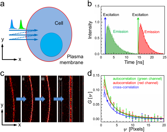
We report pulsed interleaved excitation (PIE) based line-scanning spatial correlation spectroscopy (PIE-lsSCS), a quantitative fluorescence microscopy method for the study of dynamics in free-standing lipid bilayer membranes. Using a confocal microscope, we scan multiple lines perpendicularly through the membrane, each one laterally displaced from the previous one by several ten nanometers. Scanning through the membrane enables us to eliminate intensity fluctuations due to membrane displacements with respect to the observation volume. The diffusion of fluorescent molecules within the membrane is quantified by spatial correlation analysis, based on the fixed lag times between successive line scans. PIE affords dual-color excitation within a single line scan and avoids channel crosstalk. PIE-lsSCS data are acquired from a larger membrane region so that sampling is more efficient. Moreover, the local photon flux is reduced compared with single-point experiments, resulting in a smaller fraction of photobleached molecules for identical exposure times. This is helpful for precise measurements on live cells and tissues. We have evaluated the method with experiments on fluorescently labeled giant unilamellar vesicles (GUVs) and membrane-stained live cells.
中文翻译:

基于脉冲交错激发的线扫描空间相关光谱 (PIE-lsSCS)。
我们报告了基于脉冲交错激发(PIE)的线扫描空间相关光谱(PIE-lsSCS),这是一种用于研究独立式脂质双层膜动力学的定量荧光显微镜方法。使用共焦显微镜,我们垂直穿过膜扫描多条线,每条线与前一条线横向位移几十纳米。通过膜扫描使我们能够消除由于膜相对于观察体积的位移而引起的强度波动。基于连续线扫描之间的固定滞后时间,通过空间相关分析来量化膜内荧光分子的扩散。 PIE 在单线扫描内提供双色激发并避免通道串扰。 PIE-lsSCS 数据是从更大的膜区域获取的,因此采样效率更高。此外,与单点实验相比,局部光子通量减少,导致相同曝光时间下光漂白分子的比例更小。这有助于精确测量活细胞和组织。我们通过荧光标记的巨型单层囊泡(GUV)和膜染色的活细胞的实验评估了该方法。
更新日期:2018-11-13
中文翻译:

基于脉冲交错激发的线扫描空间相关光谱 (PIE-lsSCS)。
我们报告了基于脉冲交错激发(PIE)的线扫描空间相关光谱(PIE-lsSCS),这是一种用于研究独立式脂质双层膜动力学的定量荧光显微镜方法。使用共焦显微镜,我们垂直穿过膜扫描多条线,每条线与前一条线横向位移几十纳米。通过膜扫描使我们能够消除由于膜相对于观察体积的位移而引起的强度波动。基于连续线扫描之间的固定滞后时间,通过空间相关分析来量化膜内荧光分子的扩散。 PIE 在单线扫描内提供双色激发并避免通道串扰。 PIE-lsSCS 数据是从更大的膜区域获取的,因此采样效率更高。此外,与单点实验相比,局部光子通量减少,导致相同曝光时间下光漂白分子的比例更小。这有助于精确测量活细胞和组织。我们通过荧光标记的巨型单层囊泡(GUV)和膜染色的活细胞的实验评估了该方法。


 京公网安备 11010802027423号