当前位置:
X-MOL 学术
›
Nat. Methods
›
论文详情
Our official English website, www.x-mol.net, welcomes your
feedback! (Note: you will need to create a separate account there.)
Nanobody immunostaining for correlated light and electron microscopy with preservation of ultrastructure.
Nature Methods ( IF 36.1 ) Pub Date : 2018-11-05 , DOI: 10.1038/s41592-018-0177-x Tao Fang 1 , Xiaotang Lu 2 , Daniel Berger 2 , Christina Gmeiner 2 , Julia Cho 3 , Richard Schalek 2 , Hidde Ploegh 1 , Jeff Lichtman 2
中文翻译:

纳米抗体免疫染色,用于相关的光学和电子显微镜检查,并保留超微结构。
更新日期:2018-12-10
Nature Methods ( IF 36.1 ) Pub Date : 2018-11-05 , DOI: 10.1038/s41592-018-0177-x Tao Fang 1 , Xiaotang Lu 2 , Daniel Berger 2 , Christina Gmeiner 2 , Julia Cho 3 , Richard Schalek 2 , Hidde Ploegh 1 , Jeff Lichtman 2
Affiliation

|
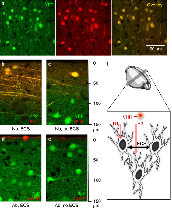
Morphological and molecular characteristics determine the function of biological tissues. Attempts to combine immunofluorescence and electron microscopy invariably compromise the quality of the ultrastructure of tissue sections. We developed NATIVE, a correlated light and electron microscopy approach that preserves ultrastructure while showing the locations of multiple molecular moieties, even deep within tissues. This technique allowed the large-scale 3D reconstruction of a volume of mouse hippocampal CA3 tissue at nanometer resolution.
中文翻译:

纳米抗体免疫染色,用于相关的光学和电子显微镜检查,并保留超微结构。
形态和分子特征决定了生物组织的功能。尝试将免疫荧光和电子显微镜相结合始终会损害组织切片超微结构的质量。我们开发了NATIVE,这是一种与光电子显微镜相关的方法,可以保留超微结构,同时显示多个分子部分的位置,甚至在组织内部。这项技术可以纳米级分辨率对小鼠海马CA3组织进行大规模3D重建。


 京公网安备 11010802027423号