Our official English website, www.x-mol.net, welcomes your
feedback! (Note: you will need to create a separate account there.)
Intravital imaging with two-photon microscopy reveals cellular dynamics in the ischeamia-reperfused rat heart.
Scientific Reports ( IF 3.8 ) Pub Date : 2018-Oct-30 , DOI: 10.1038/s41598-018-34295-w Ryohei Matsuura 1 , Shigeru Miyagawa 1 , Satsuki Fukushima 1 , Takasumi Goto 1 , Akima Harada 1 , Yuri Shimozaki 2 , Kazumasa Yamaki 2 , Sho Sanami 2 , Junichi Kikuta 3 , Masaru Ishii 3 , Yoshiki Sawa 1
Scientific Reports ( IF 3.8 ) Pub Date : 2018-Oct-30 , DOI: 10.1038/s41598-018-34295-w Ryohei Matsuura 1 , Shigeru Miyagawa 1 , Satsuki Fukushima 1 , Takasumi Goto 1 , Akima Harada 1 , Yuri Shimozaki 2 , Kazumasa Yamaki 2 , Sho Sanami 2 , Junichi Kikuta 3 , Masaru Ishii 3 , Yoshiki Sawa 1
Affiliation

|
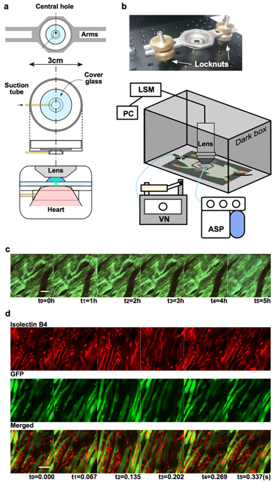
Recent advances in intravital microscopy have provided insight into dynamic biological events at the cellular level in both healthy and pathological tissue. However, real-time in vivo cellular imaging of the beating heart has not been fully established, mainly due to the difficulty of obtaining clear images through cycles of cardiac and respiratory motion. Here we report the successful recording of clear in vivo moving images of the beating rat heart by two-photon microscopy facilitated by cardiothoracic surgery and a novel cardiac stabiliser. Subcellular dynamics of the major cardiac components including the myocardium and its subcellular structures (i.e., nuclei and myofibrils) and mitochondrial distribution in cardiac myocytes were visualised for 4-5 h in green fluorescent protein-expressing transgenic Lewis rats at 15 frames/s. We also observed ischaemia/reperfusion (I/R) injury-induced suppression of the contraction/relaxation cycle and the consequent increase in cell permeability and leukocyte accumulation in cardiac tissue. I/R injury was induced in other transgenic mouse lines to further clarify the biological events in cardiac tissue. This imaging system can serve as an alternative modality for real time monitoring in animal models and cardiological drug screening, and can contribute to the development of more effective treatments for cardiac diseases.
中文翻译:

双光子显微镜的活体成像揭示了缺血再灌注大鼠心脏的细胞动力学。
活体显微镜的最新进展使我们能够深入了解健康和病理组织中细胞水平的动态生物事件。然而,跳动心脏的实时体内细胞成像尚未完全建立,主要是由于难以通过心脏和呼吸运动周期获得清晰的图像。在这里,我们报告了在心胸外科手术和新型心脏稳定器的帮助下,通过双光子显微镜成功记录了跳动的大鼠心脏的清晰体内运动图像。在表达绿色荧光蛋白的转基因 Lewis 大鼠中,以 15 帧/秒的速度观察心肌细胞中主要心脏成分(包括心肌及其亚细胞结构(即细胞核和肌原纤维))和线粒体分布的亚细胞动力学 4-5 小时。我们还观察到缺血/再灌注(I/R)损伤引起的收缩/舒张周期抑制,以及随之而来的心脏组织中细胞通透性和白细胞积累的增加。在其他转基因小鼠品系中诱导 I/R 损伤,以进一步阐明心脏组织中的生物学事件。该成像系统可以作为动物模型实时监测和心脏病药物筛选的替代方式,并有助于开发更有效的心脏病治疗方法。
更新日期:2018-10-31
中文翻译:

双光子显微镜的活体成像揭示了缺血再灌注大鼠心脏的细胞动力学。
活体显微镜的最新进展使我们能够深入了解健康和病理组织中细胞水平的动态生物事件。然而,跳动心脏的实时体内细胞成像尚未完全建立,主要是由于难以通过心脏和呼吸运动周期获得清晰的图像。在这里,我们报告了在心胸外科手术和新型心脏稳定器的帮助下,通过双光子显微镜成功记录了跳动的大鼠心脏的清晰体内运动图像。在表达绿色荧光蛋白的转基因 Lewis 大鼠中,以 15 帧/秒的速度观察心肌细胞中主要心脏成分(包括心肌及其亚细胞结构(即细胞核和肌原纤维))和线粒体分布的亚细胞动力学 4-5 小时。我们还观察到缺血/再灌注(I/R)损伤引起的收缩/舒张周期抑制,以及随之而来的心脏组织中细胞通透性和白细胞积累的增加。在其他转基因小鼠品系中诱导 I/R 损伤,以进一步阐明心脏组织中的生物学事件。该成像系统可以作为动物模型实时监测和心脏病药物筛选的替代方式,并有助于开发更有效的心脏病治疗方法。


 京公网安备 11010802027423号