当前位置:
X-MOL 学术
›
Biotechnol. J.
›
论文详情
Our official English website, www.x-mol.net, welcomes your
feedback! (Note: you will need to create a separate account there.)
Cell‐Nanoparticle Interactions at (Sub)–Nanometer Resolution Analyzed by Electron Microscopy and Correlative Coherent Anti‐Stokes Raman Scattering
Biotechnology Journal ( IF 3.2 ) Pub Date : 2018-11-23 , DOI: 10.1002/biot.201800413 Jukka Saarinen 1 , Friederike Gütter 2 , Mervi Lindman 3 , Mikael Agopov 1 , Sara J. Fraser‐Miller 1, 4 , Regina Scherließ 2 , Eija Jokitalo 3 , Hélder A. Santos 5 , Leena Peltonen 1 , Antti Isomäki 6 , Clare J. Strachan 1
Biotechnology Journal ( IF 3.2 ) Pub Date : 2018-11-23 , DOI: 10.1002/biot.201800413 Jukka Saarinen 1 , Friederike Gütter 2 , Mervi Lindman 3 , Mikael Agopov 1 , Sara J. Fraser‐Miller 1, 4 , Regina Scherließ 2 , Eija Jokitalo 3 , Hélder A. Santos 5 , Leena Peltonen 1 , Antti Isomäki 6 , Clare J. Strachan 1
Affiliation

|
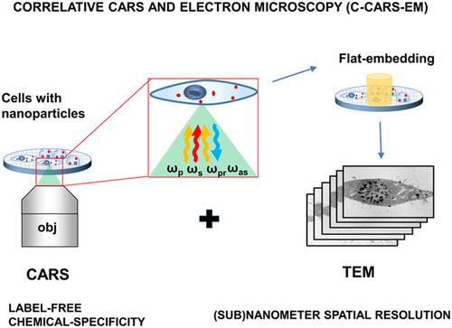
A wide variety of nanoparticles are playing an increasingly important role in drug delivery. Label‐free imaging techniques are especially desirable to follow the cellular uptake and intracellular fate of nanoparticles. The combined correlative use of different techniques, each with unique advantages, facilitates more detailed investigation about such interactions. The synergistic use of correlative coherent anti‐Stokes Raman scattering and electron microscopy (C‐CARS‐EM) imaging offers label‐free, chemically‐specific, and (sub)‐nanometer spatial resolution for studying nanoparticle uptake into cells as demonstrated in the current study. Coherent anti‐Stokes Raman scattering (CARS) microscopy offers chemically‐specific (sub)micron spatial resolution imaging without fluorescent labels while transmission electron microscopy (TEM) offers (sub)‐nanometer scale spatial resolution and thus visualization of precise nanoparticle localization at the sub‐cellular level. This proof‐of‐concept imaging platform with unlabeled drug nanocrystals and macrophage cells revealed good colocalization between the CARS signal and electron dense nanocrystals in TEM images. The correlative TEM images revealed subcellular localization of nanocrystals inside membrane bound vesicles, showing multivesicular body (MVB)−like morphology typical for late endosomes (LEs), endolysosomes, and phagolysosomes. C‐CARS‐EM imaging has much potential to study the interactions between a wide range of nanoparticles and cells with high precision and confidence.
中文翻译:

通过电子显微镜和相关相干反斯托克斯拉曼散射分析的(亚)-纳米分辨率下的细胞-纳米粒子相互作用
各种各样的纳米颗粒在药物递送中起着越来越重要的作用。跟随纳米颗粒的细胞摄取和细胞内命运,无标记成像技术尤为可取。不同技术的组合相关用法的使用,每个都有独特的优势,有助于更详细地研究此类交互。相关相干反斯托克斯拉曼散射和电子显微镜(C‐CARS‐EM)成像的协同使用可提供无标签,化学特异性和(亚)纳米级的空间分辨率,用于研究纳米粒子摄入细胞的能力,如当前所证明的那样学习。相干抗斯托克斯拉曼散射(CARS)显微镜可提供化学特异性的(亚)微米空间分辨率成像,而无需荧光标记,而透射电子显微镜(TEM)可提供(亚)纳米级的空间分辨率,从而可视化亚纳米级的精确纳米颗粒定位细胞水平。这个带有未标记药物纳米晶体和巨噬细胞的概念验证成像平台显示出TEM图像中CARS信号和电子致密纳米晶体之间的良好共定位。相关的TEM图像显示了膜结合囊泡内的纳米晶体的亚细胞定位,显示了晚期内体(LEs),内体溶酶体和吞噬体的典型多囊体(MVB)样形态。
更新日期:2019-02-26
中文翻译:

通过电子显微镜和相关相干反斯托克斯拉曼散射分析的(亚)-纳米分辨率下的细胞-纳米粒子相互作用
各种各样的纳米颗粒在药物递送中起着越来越重要的作用。跟随纳米颗粒的细胞摄取和细胞内命运,无标记成像技术尤为可取。不同技术的组合相关用法的使用,每个都有独特的优势,有助于更详细地研究此类交互。相关相干反斯托克斯拉曼散射和电子显微镜(C‐CARS‐EM)成像的协同使用可提供无标签,化学特异性和(亚)纳米级的空间分辨率,用于研究纳米粒子摄入细胞的能力,如当前所证明的那样学习。相干抗斯托克斯拉曼散射(CARS)显微镜可提供化学特异性的(亚)微米空间分辨率成像,而无需荧光标记,而透射电子显微镜(TEM)可提供(亚)纳米级的空间分辨率,从而可视化亚纳米级的精确纳米颗粒定位细胞水平。这个带有未标记药物纳米晶体和巨噬细胞的概念验证成像平台显示出TEM图像中CARS信号和电子致密纳米晶体之间的良好共定位。相关的TEM图像显示了膜结合囊泡内的纳米晶体的亚细胞定位,显示了晚期内体(LEs),内体溶酶体和吞噬体的典型多囊体(MVB)样形态。


 京公网安备 11010802027423号