Progress in Retinal and Eye Research ( IF 18.6 ) Pub Date : 2018-10-10 , DOI: 10.1016/j.preteyeres.2018.10.001 Nicola Quinn , Lajos Csincsik , Erin Flynn , Christine A. Curcio , Szilard Kiss , SriniVas R. Sadda , Ruth Hogg , Tunde Peto , Imre Lengyel

|
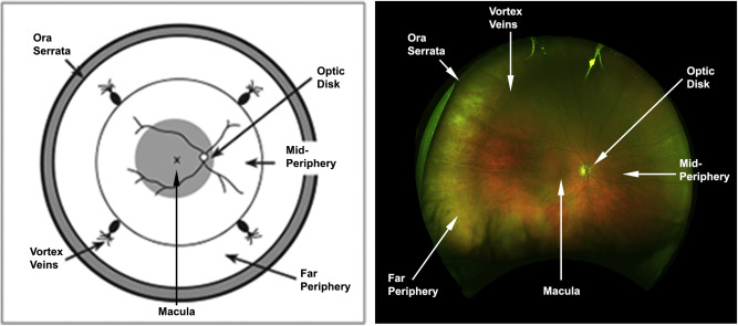
Recent developments in imaging technologies now allow the documentation, qualitative and quantitative evaluation of peripheral retinal lesions. As wide field retinal imaging, capturing both the central and peripheral retina up to 200° eccentricity, is becoming readily available the question is: what is it that we gain by imaging the periphery? Based on accumulating evidence it is clear that findings in the periphery do not always associate to those observed in the posterior pole. However, the newly acquired information may provide useful clues to previously unrecognised disease features and may facilitate more accurate disease prognostication. In this review, we explore the anatomy and physiology of the peripheral retina, focusing on how it differs from the posterior pole, recount the history of peripheral retinal imaging, describe various peripheral retinal lesions and evaluate the overall relevance of peripheral retinal findings to different diseases.
中文翻译:

可视化周围视网膜的临床意义
成像技术的最新发展现在允许对周围视网膜病变进行记录,定性和定量评估。随着捕获中心和周围视网膜高达200°偏心距的广角视网膜成像变得越来越容易获得,问题是:通过对外围进行成像,我们可以获得什么?基于积累的证据,很明显,周围的发现并不总是与在后极观察到的有关。但是,新获得的信息可能会为以前无法识别的疾病特征提供有用的线索,并可能有助于更准确的疾病预后。在这篇综述中,我们探讨了周围视网膜的解剖学和生理学,着重于它与后极的区别,叙述了周围视网膜成像的历史,


 京公网安备 11010802027423号