当前位置:
X-MOL 学术
›
Nat. Commun.
›
论文详情
Our official English website, www.x-mol.net, welcomes your
feedback! (Note: you will need to create a separate account there.)
Spatially-resolved fluorescence-detected two-dimensional electronic spectroscopy probes varying excitonic structure in photosynthetic bacteria.
Nature Communications ( IF 14.7 ) Pub Date : 2018-10-11 , DOI: 10.1038/s41467-018-06619-x Vivek Tiwari , Yassel Acosta Matutes , Alastair T. Gardiner , Thomas L. C. Jansen , Richard J. Cogdell , Jennifer P. Ogilvie
Nature Communications ( IF 14.7 ) Pub Date : 2018-10-11 , DOI: 10.1038/s41467-018-06619-x Vivek Tiwari , Yassel Acosta Matutes , Alastair T. Gardiner , Thomas L. C. Jansen , Richard J. Cogdell , Jennifer P. Ogilvie

|
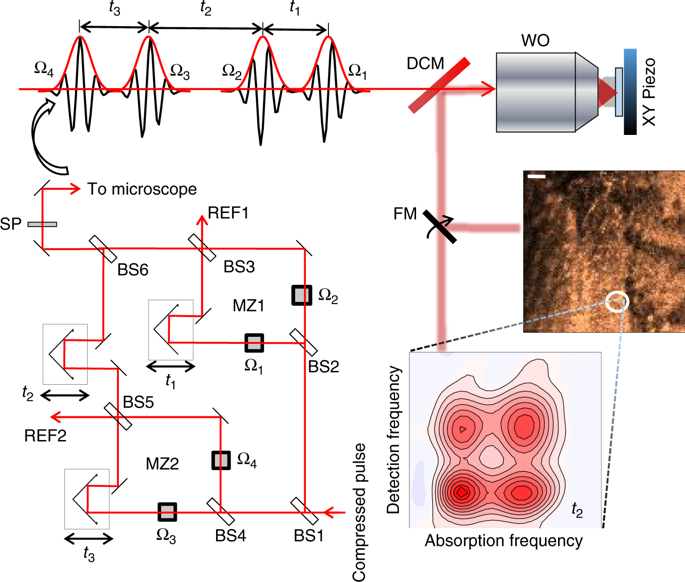
Conventional implementations of two-dimensional electronic spectroscopy typically spatially average over ~1010 chromophores spread over ~104 micron square area, limiting their ability to characterize spatially heterogeneous samples. Here we present a variation of two-dimensional electronic spectroscopy that is capable of mapping spatially varying differences in excitonic structure, with sensitivity orders of magnitude better than conventional spatially-averaged electronic spectroscopies. The approach performs fluorescence-detection-based fully collinear two-dimensional electronic spectroscopy in a microscope, combining femtosecond time-resolution, sub-micron spatial resolution, and the sensitivity of fluorescence detection. We demonstrate the approach on a mixture of photosynthetic bacteria that are known to exhibit variations in electronic structure with growth conditions. Spatial variations in the constitution of mixed bacterial colonies manifests as spatially varying peak intensities in the measured two-dimensional contour maps, which exhibit distinct diagonal and cross-peaks that reflect differences in the excitonic structure of the bacterial proteins.
中文翻译:

空间分辨荧光检测二维电子光谱探针可探测光合细菌中激子结构的变化。
二维电子光谱的常规实现通常在〜10 10个发色团上分布在〜10 4个上的空间平均微米见方的面积,限制了它们表征空间异质样品的能力。在这里,我们介绍了一种二维电子光谱的变体,它能够映射激子结构在空间上变化的差异,其灵敏度比传统的空间平均电子光谱要好几个数量级。该方法在显微镜中执行了基于荧光检测的全共线二维电子光谱,结合了飞秒时间分辨率,亚微米空间分辨率和荧光检测的灵敏度。我们展示了一种对光合细菌混合物的处理方法,已知该混合物在生长条件下会显示出电子结构的变化。
更新日期:2018-10-11
中文翻译:

空间分辨荧光检测二维电子光谱探针可探测光合细菌中激子结构的变化。
二维电子光谱的常规实现通常在〜10 10个发色团上分布在〜10 4个上的空间平均微米见方的面积,限制了它们表征空间异质样品的能力。在这里,我们介绍了一种二维电子光谱的变体,它能够映射激子结构在空间上变化的差异,其灵敏度比传统的空间平均电子光谱要好几个数量级。该方法在显微镜中执行了基于荧光检测的全共线二维电子光谱,结合了飞秒时间分辨率,亚微米空间分辨率和荧光检测的灵敏度。我们展示了一种对光合细菌混合物的处理方法,已知该混合物在生长条件下会显示出电子结构的变化。


 京公网安备 11010802027423号