当前位置:
X-MOL 学术
›
J. Biophotonics
›
论文详情
Our official English website, www.x-mol.net, welcomes your
feedback! (Note: you will need to create a separate account there.)
Superparamagnetic graphene quantum dot as a dual-modality contrast agent for confocal fluorescence microscopy and magnetomotive optical coherence tomography.
Journal of Biophotonics ( IF 2.0 ) Pub Date : 2018-10-21 , DOI: 10.1002/jbio.201800219 Wei Li 1 , Wenxing Song 2 , Biqiong Chen 3 , Stephen J Matcher 1
Journal of Biophotonics ( IF 2.0 ) Pub Date : 2018-10-21 , DOI: 10.1002/jbio.201800219 Wei Li 1 , Wenxing Song 2 , Biqiong Chen 3 , Stephen J Matcher 1
Affiliation
|
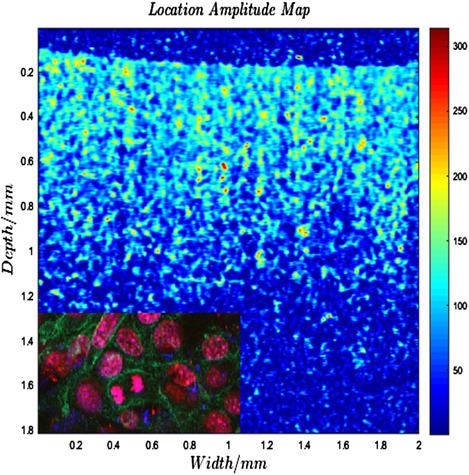
A magnetic graphene quantum dot (MGQD) nanoparticle, synthesized by hydrothermally reducing and cutting graphene oxide‐iron oxide sheet, was demonstrated to possess the capabilities of simultaneous confocal fluorescence and magnetomotive optical coherence tomography (MMOCT) imaging. This MGQD shows low toxicity, significant tunable blue fluorescence and superparamagnetism, which can thus be used as a dual‐modality contrast agent for confocal fluorescence microscopy (CFM) and MMOCT. The feasibility of applying MGQD as a tracer of cells is shown by imaging and visualizing MGQD labeled cells using CFM and our in‐house MMOCT. Since MMOCT and CFM can offer anatomical structure and intracellular details, respectively, the MGQD for cell tracking could provide a more comprehensive diagnosis.
中文翻译:

超顺磁性石墨烯量子点作为共模荧光显微镜和磁动力光学相干断层扫描的双峰型造影剂。
通过水热还原和切割氧化石墨烯-氧化铁片合成的磁性石墨烯量子点(MGQD)纳米粒子具有同步共聚焦荧光和磁动光学相干层析成像(MMOCT)成像的功能。该MGQD显示出低毒性,明显的可调谐蓝色荧光和超顺磁性,因此可用作共焦荧光显微镜(CFM)和MMOCT的双峰造影剂。通过使用CFM和我们的内部MMOCT对MGQD标记的细胞进行成像和可视化,可以证明将MGQD用作细胞示踪剂的可行性。由于MMOCT和CFM可以分别提供解剖结构和细胞内细节,因此用于细胞跟踪的MGQD可以提供更全面的诊断。
更新日期:2018-10-21
中文翻译:
超顺磁性石墨烯量子点作为共模荧光显微镜和磁动力光学相干断层扫描的双峰型造影剂。
通过水热还原和切割氧化石墨烯-氧化铁片合成的磁性石墨烯量子点(MGQD)纳米粒子具有同步共聚焦荧光和磁动光学相干层析成像(MMOCT)成像的功能。该MGQD显示出低毒性,明显的可调谐蓝色荧光和超顺磁性,因此可用作共焦荧光显微镜(CFM)和MMOCT的双峰造影剂。通过使用CFM和我们的内部MMOCT对MGQD标记的细胞进行成像和可视化,可以证明将MGQD用作细胞示踪剂的可行性。由于MMOCT和CFM可以分别提供解剖结构和细胞内细节,因此用于细胞跟踪的MGQD可以提供更全面的诊断。


 京公网安备 11010802027423号