Our official English website, www.x-mol.net, welcomes your
feedback! (Note: you will need to create a separate account there.)
Cellulose Microfibril Formation by Surface-Tethered Cellulose Synthase Enzymes
ACS Nano ( IF 15.8 ) Pub Date : 2016-01-28 00:00:00 , DOI: 10.1021/acsnano.5b05648 Snehasish Basu 1 , Okako Omadjela 2 , David Gaddes 3 , Srinivas Tadigadapa 3 , Jochen Zimmer 2 , Jeffrey M. Catchmark 1
ACS Nano ( IF 15.8 ) Pub Date : 2016-01-28 00:00:00 , DOI: 10.1021/acsnano.5b05648 Snehasish Basu 1 , Okako Omadjela 2 , David Gaddes 3 , Srinivas Tadigadapa 3 , Jochen Zimmer 2 , Jeffrey M. Catchmark 1
Affiliation

|
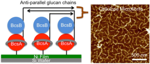
Cellulose microfibrils are pseudocrystalline arrays of cellulose chains that are synthesized by cellulose synthases. The enzymes are organized into large membrane-embedded complexes in which each enzyme likely synthesizes and secretes a β-(1→4) glucan. The relationship between the organization of the enzymes in these complexes and cellulose crystallization has not been explored. To better understand this relationship, we used atomic force microscopy to visualize cellulose microfibril formation from nickel-film-immobilized bacterial cellulose synthase enzymes (BcsA-Bs), which in standard solution only form amorphous cellulose from monomeric BcsA-B complexes. Fourier transform infrared spectroscopy and X-ray diffraction techniques show that surface-tethered BcsA-Bs synthesize highly crystalline cellulose II in the presence of UDP-Glc, the allosteric activator cyclic-di-GMP, as well as magnesium. The cellulose II cross section/diameter and the crystal size and crystallinity depend on the surface density of tethered enzymes as well as the overall concentration of substrates. Our results provide the correlation between cellulose microfibril formation and the spatial organization of cellulose synthases.
中文翻译:

通过表面束缚的纤维素合酶的纤维素微纤维形成。
纤维素微原纤维是由纤维素合成酶合成的纤维素链的假晶阵列。这些酶被组织成大型的膜嵌入复合物,其中每种酶都可能合成并分泌β-(1→4)葡聚糖。这些复合物中酶的组织与纤维素结晶之间的关系尚未探索。为了更好地理解这种关系,我们使用原子力显微镜观察了固定有镍膜的细菌纤维素合成酶(BcsA-Bs)形成的纤维素微原纤维,该酶在标准溶液中仅由单体BcsA-B复合物形成无定形纤维素。傅立叶变换红外光谱和X射线衍射技术表明,在UDP-Glc的存在下,表面束缚的BcsA-Bs可以合成高度结晶的纤维素II,变构活化剂环二GMP以及镁。纤维素II的横截面/直径以及晶体的大小和结晶度取决于拴系酶的表面密度以及底物的总浓度。我们的结果提供了纤维素微原纤维形成与纤维素合成酶的空间组织之间的相关性。
更新日期:2016-01-28
中文翻译:

通过表面束缚的纤维素合酶的纤维素微纤维形成。
纤维素微原纤维是由纤维素合成酶合成的纤维素链的假晶阵列。这些酶被组织成大型的膜嵌入复合物,其中每种酶都可能合成并分泌β-(1→4)葡聚糖。这些复合物中酶的组织与纤维素结晶之间的关系尚未探索。为了更好地理解这种关系,我们使用原子力显微镜观察了固定有镍膜的细菌纤维素合成酶(BcsA-Bs)形成的纤维素微原纤维,该酶在标准溶液中仅由单体BcsA-B复合物形成无定形纤维素。傅立叶变换红外光谱和X射线衍射技术表明,在UDP-Glc的存在下,表面束缚的BcsA-Bs可以合成高度结晶的纤维素II,变构活化剂环二GMP以及镁。纤维素II的横截面/直径以及晶体的大小和结晶度取决于拴系酶的表面密度以及底物的总浓度。我们的结果提供了纤维素微原纤维形成与纤维素合成酶的空间组织之间的相关性。


 京公网安备 11010802027423号