当前位置:
X-MOL 学术
›
Acta Biomater.
›
论文详情
Our official English website, www.x-mol.net, welcomes your
feedback! (Note: you will need to create a separate account there.)
Bone matrix development in steroid-induced osteoporosis is associated with a consistently reduced fibrillar stiffness linked to altered bone mineral quality.
Acta Biomaterialia ( IF 9.4 ) Pub Date : 2018-06-15 , DOI: 10.1016/j.actbio.2018.05.053 L Xi 1 , P De Falco 2 , E Barbieri 3 , A Karunaratne 4 , L Bentley 5 , C T Esapa 6 , N J Terrill 7 , S D M Brown 5 , R D Cox 5 , G R Davis 8 , N M Pugno 9 , R V Thakker 6 , H S Gupta 10
Acta Biomaterialia ( IF 9.4 ) Pub Date : 2018-06-15 , DOI: 10.1016/j.actbio.2018.05.053 L Xi 1 , P De Falco 2 , E Barbieri 3 , A Karunaratne 4 , L Bentley 5 , C T Esapa 6 , N J Terrill 7 , S D M Brown 5 , R D Cox 5 , G R Davis 8 , N M Pugno 9 , R V Thakker 6 , H S Gupta 10
Affiliation
|
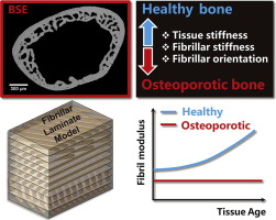
Glucocorticoid-induced osteoporosis (GIOP) is a major secondary form of osteoporosis, with the fracture risk significantly elevated - at similar levels of bone mineral density - in patients taking glucocorticoids compared with non-users. The adverse bone structural changes at multiple hierarchical levels in GIOP, and their mechanistic consequences leading to reduced load-bearing capacity, are not clearly understood. Here we combine experimental X-ray nanoscale mechanical imaging with analytical modelling of the bone matrix mechanics to determine mechanisms causing bone material quality deterioration during development of GIOP. In situ synchrotron small-angle X-ray diffraction combined with tensile testing was used to measure nanoscale deformation mechanisms in a murine model of GIOP, due to a corticotrophin-releasing hormone promoter mutation, at multiple ages (8-, 12-, 24- and 36 weeks), complemented by quantitative micro-computed tomography and backscattered electron imaging to determine mineral concentrations. We develop a two-level hierarchical model of the bone matrix (mineralized fibril and lamella) to predict fibrillar mechanical response as a function of architectural parameters of the mineralized matrix. The fibrillar elastic modulus of GIOP-bone is lower than healthy bone throughout development, and nearly constant in time, in contrast to the progressively increasing stiffness in healthy bone. The lower mineral platelet aspect ratio value for GIOP compared to healthy bone in the multiscale model can explain the fibrillar deformation. Consistent with this result, independent measurement of mineral platelet lengths from wide-angle X-ray diffraction finds a shorter mineral platelet length in GIOP. Our results show how lowered mineralization combined with altered mineral nanostructure in GIOP leads to lowered mechanical competence.
SIGNIFICANCE STATEMENT
Increased fragility in musculoskeletal disorders like osteoporosis are believed to arise due to alterations in bone structure at multiple length-scales from the organ down to the supramolecular-level, where collagen molecules and elongated mineral nanoparticles form stiff fibrils. However, the nature of these molecular-level alterations are not known. Here we used X-ray scattering to determine both how bone fibrils deform in secondary osteoporosis, as well as how the fibril orientation and mineral nanoparticle structure changes. We found that osteoporotic fibrils become less stiff both because the mineral nanoparticles became shorter and less efficient at transferring load from collagen, and because the fibrils are more randomly oriented. These results will help in the design of new composite musculoskeletal implants for bone repair.
中文翻译:

类固醇引起的骨质疏松症中的骨基质发育与纤维硬度持续降低有关,而纤维硬度持续降低,而纤维硬度又与骨矿物质质量的改变有关。
糖皮质激素引起的骨质疏松症 (GIOP) 是骨质疏松症的一种主要继发形式,与未使用糖皮质激素的患者相比,在骨密度相似的情况下,服用糖皮质激素的患者骨折风险显着升高。GIOP 中多个层次的不利骨结构变化及其导致承载能力降低的机制后果尚不清楚。在这里,我们将实验性 X 射线纳米级机械成像与骨基质力学的分析模型相结合,以确定在 GIOP 发育过程中导致骨材料质量恶化的机制。原位同步加速器小角度 X 射线衍射与拉伸测试相结合,用于测量 GIOP 小鼠模型中由于促肾上腺皮质激素释放激素启动子突变而导致的多个年龄(8、12、24 岁)的纳米级变形机制。和 36 周),并辅以定量微型计算机断层扫描和背散射电子成像来确定矿物质浓度。我们开发了骨基质(矿化原纤维和板层)的两级分层模型,以预测纤维机械响应作为矿化基质结构参数的函数。GIOP骨的纤维弹性模量在整个发育过程中低于健康骨,并且随时间几乎恒定,这与健康骨中逐渐增加的刚度相反。在多尺度模型中,与健康骨骼相比,GIOP 的矿物质血小板长宽比值较低,可以解释纤维变形。与该结果一致,通过广角 X 射线衍射对矿物片长度进行独立测量,发现 GIOP 中的矿物片长度较短。我们的结果表明,GIOP 中矿化度的降低与矿物纳米结构的改变是如何导致机械性能降低的。意义声明 骨质疏松症等肌肉骨骼疾病的脆性增加被认为是由于从器官到超分子水平的多个长度尺度的骨结构改变而引起的,其中胶原蛋白分子和细长的矿物纳米颗粒形成坚硬的原纤维。然而,这些分子水平改变的性质尚不清楚。在这里,我们使用 X 射线散射来确定继发性骨质疏松症中骨原纤维如何变形,以及原纤维方向和矿物纳米颗粒结构如何变化。我们发现,骨质疏松原纤维变得不那么僵硬,这既是因为矿物纳米颗粒变得更短,传递胶原蛋白负载的效率较低,也是因为原纤维的方向更加随机。这些结果将有助于设计用于骨修复的新型复合肌肉骨骼植入物。
更新日期:2018-06-15
中文翻译:
类固醇引起的骨质疏松症中的骨基质发育与纤维硬度持续降低有关,而纤维硬度持续降低,而纤维硬度又与骨矿物质质量的改变有关。
糖皮质激素引起的骨质疏松症 (GIOP) 是骨质疏松症的一种主要继发形式,与未使用糖皮质激素的患者相比,在骨密度相似的情况下,服用糖皮质激素的患者骨折风险显着升高。GIOP 中多个层次的不利骨结构变化及其导致承载能力降低的机制后果尚不清楚。在这里,我们将实验性 X 射线纳米级机械成像与骨基质力学的分析模型相结合,以确定在 GIOP 发育过程中导致骨材料质量恶化的机制。原位同步加速器小角度 X 射线衍射与拉伸测试相结合,用于测量 GIOP 小鼠模型中由于促肾上腺皮质激素释放激素启动子突变而导致的多个年龄(8、12、24 岁)的纳米级变形机制。和 36 周),并辅以定量微型计算机断层扫描和背散射电子成像来确定矿物质浓度。我们开发了骨基质(矿化原纤维和板层)的两级分层模型,以预测纤维机械响应作为矿化基质结构参数的函数。GIOP骨的纤维弹性模量在整个发育过程中低于健康骨,并且随时间几乎恒定,这与健康骨中逐渐增加的刚度相反。在多尺度模型中,与健康骨骼相比,GIOP 的矿物质血小板长宽比值较低,可以解释纤维变形。与该结果一致,通过广角 X 射线衍射对矿物片长度进行独立测量,发现 GIOP 中的矿物片长度较短。我们的结果表明,GIOP 中矿化度的降低与矿物纳米结构的改变是如何导致机械性能降低的。意义声明 骨质疏松症等肌肉骨骼疾病的脆性增加被认为是由于从器官到超分子水平的多个长度尺度的骨结构改变而引起的,其中胶原蛋白分子和细长的矿物纳米颗粒形成坚硬的原纤维。然而,这些分子水平改变的性质尚不清楚。在这里,我们使用 X 射线散射来确定继发性骨质疏松症中骨原纤维如何变形,以及原纤维方向和矿物纳米颗粒结构如何变化。我们发现,骨质疏松原纤维变得不那么僵硬,这既是因为矿物纳米颗粒变得更短,传递胶原蛋白负载的效率较低,也是因为原纤维的方向更加随机。这些结果将有助于设计用于骨修复的新型复合肌肉骨骼植入物。


 京公网安备 11010802027423号