Our official English website, www.x-mol.net, welcomes your
feedback! (Note: you will need to create a separate account there.)
On the Formation and Morphology of Lipid Nanoparticles Containing Ionizable Cationic Lipids and siRNA
ACS Nano ( IF 15.8 ) Pub Date : 2018-04-03 00:00:00 , DOI: 10.1021/acsnano.8b01516 Jayesh A. Kulkarni 1 , Maria M. Darjuan 1 , Joanne E. Mercer 2 , Sam Chen 1, 3 , Roy van der Meel 1, 4 , Jenifer L. Thewalt 2 , Yuen Yi C. Tam 1, 3 , Pieter R. Cullis 1
ACS Nano ( IF 15.8 ) Pub Date : 2018-04-03 00:00:00 , DOI: 10.1021/acsnano.8b01516 Jayesh A. Kulkarni 1 , Maria M. Darjuan 1 , Joanne E. Mercer 2 , Sam Chen 1, 3 , Roy van der Meel 1, 4 , Jenifer L. Thewalt 2 , Yuen Yi C. Tam 1, 3 , Pieter R. Cullis 1
Affiliation

|
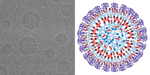
Lipid nanoparticles (LNPs) containing short interfering RNA (LNP-siRNA) and optimized ionizable cationic lipids are now clinically validated systems for silencing disease-causing genes in hepatocytes following intravenous administration. However, the mechanism of formation and certain structural features of LNP-siRNA remain obscure. These systems are formed from lipid mixtures (cationic lipid, distearoylphosphatidylcholine, cholesterol, and PEG-lipid) dissolved in ethanol that is rapidly mixed with siRNA in aqueous buffer at a pH (pH 4) where the ionizable lipid is positively charged. The resulting dispersion is then dialyzed against a normal saline buffer to remove residual ethanol and raise the pH to 7.4 (above the pKa of the cationic lipid) to produce the finished LNP-siRNA systems. Here we provide cryogenic transmission electron microscopy (cryo-TEM) and X-ray evidence that the complexes formed between siRNA and ionizable lipid at pH 4 correspond to tightly packed bilayer structures with siRNA sandwiched between closely apposed monolayers. Further, it is shown that ionizable lipid not complexed to siRNA promotes formation of very small vesicular structures at pH 4 that coalesce to form larger LNP structures with amorphous electron dense cores at pH 7.4. A mechanism of formation of LNP-siRNA systems is proposed whereby siRNA is first sandwiched between closely apposed lipid monolayers at pH 4 and subsequently trapped in these structures as the pH is raised to 7.4, whereas ionizable lipid not interacting with siRNA moves from bilayer structure to adopt an amorphous oil phase located in the center of the LNP as the pH is raised. This model is discussed in terms of previous hypotheses and potential relevance to the design of LNP-siRNA systems.
中文翻译:

含可电离阳离子脂质和siRNA的脂质纳米颗粒的形成和形貌
包含短干扰RNA(LNP-siRNA)和优化的可离子化阳离子脂质的脂质纳米颗粒(LNP)现在已成为临床验证的系统,用于在静脉内给药后沉默肝细胞中的致病基因。但是,LNP-siRNA的形成机理和某些结构特征仍然不清楚。这些系统由溶解在乙醇中的脂质混合物(阳离子脂质,二硬脂酰磷脂酰胆碱,胆固醇和PEG-脂质)形成,该脂质混合物与siRNA在pH(pH 4)的水性缓冲液中快速混合,其中可电离的脂质带正电。然后将所得的分散液在生理盐水缓冲液中透析,以去除残留的乙醇,并将pH升高至7.4(高于p K a阳离子脂质)以生产最终的LNP-siRNA系统。在这里,我们提供了低温透射电子显微镜(cryo-TEM)和X射线证据,表明在pH 4时siRNA和可电离脂质之间形成的复合物对应于紧密堆积的双层结构,其中siRNA夹在紧密排列的单分子层之间。此外,显示出不与siRNA复合的可电离的脂质促进在pH 4下非常小的囊泡结构的形成,所述小囊泡结构聚结以在pH 7.4下形成具有无定形电子致密核的更大的LNP结构。提出了形成LNP-siRNA系统的机制,其中siRNA首先在pH为4的情况下夹在紧密结合的脂质单分子层之间,然后在pH升高至7.4时被困在这些结构中,而不与siRNA相互作用的可离子化脂质则从双层结构移至在pH升高时位于LNP中心的无定形油相。根据先前的假设以及与LNP-siRNA系统设计的潜在相关性讨论了该模型。
更新日期:2018-04-03
中文翻译:

含可电离阳离子脂质和siRNA的脂质纳米颗粒的形成和形貌
包含短干扰RNA(LNP-siRNA)和优化的可离子化阳离子脂质的脂质纳米颗粒(LNP)现在已成为临床验证的系统,用于在静脉内给药后沉默肝细胞中的致病基因。但是,LNP-siRNA的形成机理和某些结构特征仍然不清楚。这些系统由溶解在乙醇中的脂质混合物(阳离子脂质,二硬脂酰磷脂酰胆碱,胆固醇和PEG-脂质)形成,该脂质混合物与siRNA在pH(pH 4)的水性缓冲液中快速混合,其中可电离的脂质带正电。然后将所得的分散液在生理盐水缓冲液中透析,以去除残留的乙醇,并将pH升高至7.4(高于p K a阳离子脂质)以生产最终的LNP-siRNA系统。在这里,我们提供了低温透射电子显微镜(cryo-TEM)和X射线证据,表明在pH 4时siRNA和可电离脂质之间形成的复合物对应于紧密堆积的双层结构,其中siRNA夹在紧密排列的单分子层之间。此外,显示出不与siRNA复合的可电离的脂质促进在pH 4下非常小的囊泡结构的形成,所述小囊泡结构聚结以在pH 7.4下形成具有无定形电子致密核的更大的LNP结构。提出了形成LNP-siRNA系统的机制,其中siRNA首先在pH为4的情况下夹在紧密结合的脂质单分子层之间,然后在pH升高至7.4时被困在这些结构中,而不与siRNA相互作用的可离子化脂质则从双层结构移至在pH升高时位于LNP中心的无定形油相。根据先前的假设以及与LNP-siRNA系统设计的潜在相关性讨论了该模型。


 京公网安备 11010802027423号