Neuropsychologia ( IF 2.0 ) Pub Date : 2018-03-30 , DOI: 10.1016/j.neuropsychologia.2018.03.036
Stephanie J. Forkel , Marco Catani
|
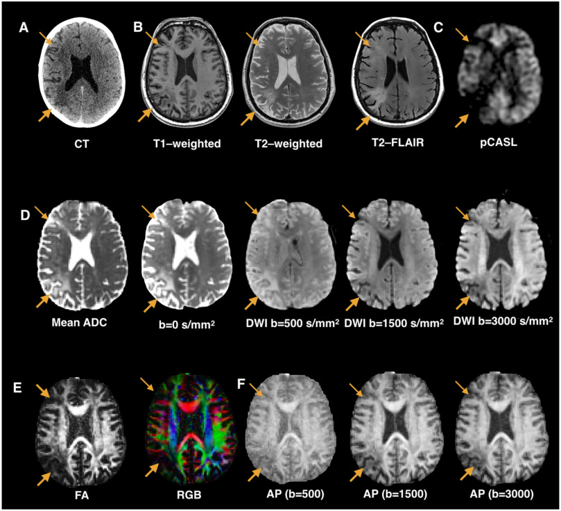
Patients with stroke offer a unique window into understanding human brain function. Mapping stroke lesions poses several challenges due to the complexity of the lesion anatomy and the mechanisms causing local and remote disruption on brain networks. In this prospective longitudinal study, we compare standard and advanced approaches to white matter lesion mapping applied to acute stroke patients with aphasia. Eighteen patients with acute left hemisphere stroke were recruited and scanned within two weeks from symptom onset. Aphasia assessment was performed at baseline and six-month follow-up. Structural and diffusion MRI contrasts indicated an area of maximum overlap in the anterior external/extreme capsule with diffusion images showing a larger overlap extending into posterior perisylvian regions. Anatomical predictors of recovery included damage to ipsilesional tracts (as shown by both structural and diffusion images) and contralesional tracts (as shown by diffusion images only). These findings indicate converging results from structural and diffusion lesion mapping methods but also clear differences between the two approaches in their ability to identify predictors of recovery outside the lesioned regions.
中文翻译:
急性中风失语症的病灶定位及其对恢复的意义
中风患者为了解人脑功能提供了一个独特的窗口。由于病变解剖结构的复杂性以及导致大脑网络局部和远程破坏的机制,绘制卒中病变图面临一些挑战。在这项前瞻性纵向研究中,我们比较了应用于急性中风失语症患者的白质病灶定位的标准方法和高级方法。在症状发作后的两周内,招募了18例急性左半球卒中患者,并对其进行了扫描。在基线和六个月的随访中进行了失语症评估。结构和弥散MRI对比显示前外部/极端囊的最大重叠区域,弥散图像显示较大的重叠延伸到后囊壁周围区域。恢复的解剖学预测指标包括对同侧道(如结构图和扩散图所示)和对侧道(如仅由扩散图所示)的损害。这些发现表明结构性和扩散性病变定位方法的结果趋同,但两种方法在识别病变区域之外的恢复预测因素的能力上也存在明显差异。


 京公网安备 11010802027423号