Our official English website, www.x-mol.net, welcomes your
feedback! (Note: you will need to create a separate account there.)
Dynamics of clusterin protein expression in the brain and plasma following experimental traumatic brain injury.
Scientific Reports ( IF 3.8 ) Pub Date : 2019-12-27 , DOI: 10.1038/s41598-019-56683-6 Shalini Das Gupta 1 , Anssi Lipponen 1 , Kaisa M A Paldanius 1 , Noora Puhakka 1 , Asla Pitkänen 1
Scientific Reports ( IF 3.8 ) Pub Date : 2019-12-27 , DOI: 10.1038/s41598-019-56683-6 Shalini Das Gupta 1 , Anssi Lipponen 1 , Kaisa M A Paldanius 1 , Noora Puhakka 1 , Asla Pitkänen 1
Affiliation

|
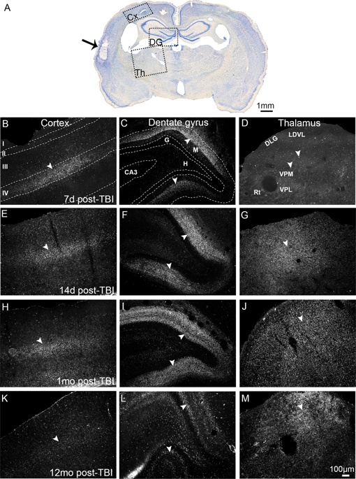
Progress in the preclinical and clinical development of neuroprotective and antiepileptogenic treatments for traumatic brain injury (TBI) necessitates the discovery of prognostic biomarkers for post-injury outcome. Our previous mRNA-seq data revealed a 1.8-2.5 fold increase in clusterin mRNA expression in lesioned brain areas in rats with lateral fluid-percussion injury (FPI)-induced TBI. On this basis, we hypothesized that TBI leads to increases in the brain levels of clusterin protein, and consequently, increased plasma clusterin levels. For evaluation, we induced TBI in adult male Sprague-Dawley rats (n = 80) by lateral FPI. We validated our mRNA-seq findings with RT-qPCR, confirming increased clusterin mRNA levels in the perilesional cortex (FC 3.3, p < 0.01) and ipsilateral thalamus (FC 2.4, p < 0.05) at 3 months post-TBI. Immunohistochemistry revealed a marked increase in extracellular clusterin protein expression in the perilesional cortex and ipsilateral hippocampus (7d to 1 month post-TBI), and ipsilateral thalamus (14d to 12 months post-TBI). In the thalamus, punctate immunoreactivity was most intense around activated microglia and mitochondria. Enzyme-linked immunoassays indicated that an acute 15% reduction, rather than an increase in plasma clusterin levels differentiated animals with TBI from sham-operated controls (AUC 0.851, p < 0.05). Our findings suggest that plasma clusterin is a candidate biomarker for acute TBI diagnosis.
中文翻译:

实验性脑外伤后脑和血浆中簇蛋白蛋白表达的动态。
创伤性脑损伤(TBI)的神经保护和抗癫痫治疗的临床前和临床开发进展,需要发现损伤后预后的生物标志物。我们以前的mRNA-seq数据显示,在患有侧向流体冲击损伤(FPI)诱导的TBI的大鼠中,病变脑区域的clusterin mRNA表达增加了1.8-2.5倍。在此基础上,我们假设TBI导致大脑中簇蛋白水平升高,从而导致血浆簇蛋白水平升高。为了进行评估,我们通过侧向FPI在成年雄性Sprague-Dawley大鼠(n = 80)中诱导了TBI。我们用RT-qPCR验证了我们的mRNA序列发现,证实了TBI后3个月,病灶周围皮层(FC 3.3,p <0.01)和同侧丘脑(FC 2.4,p <0.05)的clusterin mRNA水平增加。免疫组织化学显示,病灶周围皮层和同侧海马(TBI后7d至1个月)和同侧丘脑(TBI后14d至12个月)细胞外簇蛋白表达明显增加。在丘脑中,在活化的小胶质细胞和线粒体周围,点状免疫反应最为强烈。酶联免疫分析表明,急性TBI使动物的血浆簇蛋白水平降低了15%,而不是增加,而与假手术的对照区分开(AUC 0.851,p <0.05)。我们的发现表明血浆簇蛋白是急性TBI诊断的候选生物标志物。在丘脑中,在活化的小胶质细胞和线粒体周围,点状免疫反应最为强烈。酶联免疫分析表明,急性TBI使动物的血浆簇蛋白水平降低了15%,而不是增加,而与假手术的对照区分开(AUC 0.851,p <0.05)。我们的发现表明血浆簇蛋白是急性TBI诊断的候选生物标志物。在丘脑中,在活化的小胶质细胞和线粒体周围,点状免疫反应最为强烈。酶联免疫分析表明,急性TBI使动物的血浆簇蛋白水平降低了15%,而不是增加,而与假手术的对照区分开(AUC 0.851,p <0.05)。我们的发现表明血浆簇蛋白是急性TBI诊断的候选生物标志物。
更新日期:2019-12-27
中文翻译:

实验性脑外伤后脑和血浆中簇蛋白蛋白表达的动态。
创伤性脑损伤(TBI)的神经保护和抗癫痫治疗的临床前和临床开发进展,需要发现损伤后预后的生物标志物。我们以前的mRNA-seq数据显示,在患有侧向流体冲击损伤(FPI)诱导的TBI的大鼠中,病变脑区域的clusterin mRNA表达增加了1.8-2.5倍。在此基础上,我们假设TBI导致大脑中簇蛋白水平升高,从而导致血浆簇蛋白水平升高。为了进行评估,我们通过侧向FPI在成年雄性Sprague-Dawley大鼠(n = 80)中诱导了TBI。我们用RT-qPCR验证了我们的mRNA序列发现,证实了TBI后3个月,病灶周围皮层(FC 3.3,p <0.01)和同侧丘脑(FC 2.4,p <0.05)的clusterin mRNA水平增加。免疫组织化学显示,病灶周围皮层和同侧海马(TBI后7d至1个月)和同侧丘脑(TBI后14d至12个月)细胞外簇蛋白表达明显增加。在丘脑中,在活化的小胶质细胞和线粒体周围,点状免疫反应最为强烈。酶联免疫分析表明,急性TBI使动物的血浆簇蛋白水平降低了15%,而不是增加,而与假手术的对照区分开(AUC 0.851,p <0.05)。我们的发现表明血浆簇蛋白是急性TBI诊断的候选生物标志物。在丘脑中,在活化的小胶质细胞和线粒体周围,点状免疫反应最为强烈。酶联免疫分析表明,急性TBI使动物的血浆簇蛋白水平降低了15%,而不是增加,而与假手术的对照区分开(AUC 0.851,p <0.05)。我们的发现表明血浆簇蛋白是急性TBI诊断的候选生物标志物。在丘脑中,在活化的小胶质细胞和线粒体周围,点状免疫反应最为强烈。酶联免疫分析表明,急性TBI使动物的血浆簇蛋白水平降低了15%,而不是增加,而与假手术的对照区分开(AUC 0.851,p <0.05)。我们的发现表明血浆簇蛋白是急性TBI诊断的候选生物标志物。


 京公网安备 11010802027423号