Our official English website, www.x-mol.net, welcomes your
feedback! (Note: you will need to create a separate account there.)
Emitting/Sensitizing Ions Spatially Separated Lanthanide Nanocrystals for Visualizing Tumors Simultaneously through Up- and Down-Conversion Near-Infrared II Luminescence In Vivo.
Small ( IF 13.0 ) Pub Date : 2019-11-25 , DOI: 10.1002/smll.201905344 Yingying Li 1, 2 , Peisen Zhang 1, 2 , Haoran Ning 1, 2 , Jianfeng Zeng 3 , Yi Hou 1 , Lihong Jing 1 , Chunyan Liu 1 , Mingyuan Gao 1, 2, 3
Small ( IF 13.0 ) Pub Date : 2019-11-25 , DOI: 10.1002/smll.201905344 Yingying Li 1, 2 , Peisen Zhang 1, 2 , Haoran Ning 1, 2 , Jianfeng Zeng 3 , Yi Hou 1 , Lihong Jing 1 , Chunyan Liu 1 , Mingyuan Gao 1, 2, 3
Affiliation

|
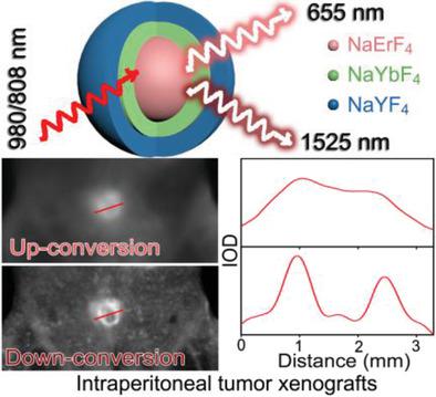
Near-infrared lights have received increasing attention regarding imaging applications owing to their large tissue penetration depth, high spatial resolution, and outstanding signal-to-noise ratio, particularly those falling in the second near-infrared window (NIR II) of biological tissues. Rare earth nanoparticles containing Er3+ ions are promising candidates to show up-conversion luminescence in the first near-infrared window (NIR I) and down-conversion luminescence in NIR II as well. However, synthesizing particles with small size and high NIR II luminescence quantum yield (QY) remains challenging. Er3+ ions are herein innovatively combined with Yb3+ ions in a NaErF4 @NaYbF4 core/shell manner instead of being codoped into NaLnF4 matrices, to maximize the concentration of Er3+ in the emitting core. After further surface coating, NaErF4 @NaYbF4 @NaYF4 core/shell/shell particles are obtained. Spectroscopy studies are carried out to show the synergistic impacts of the intermediate NaYbF4 layer and the outer NaYF4 shell. Finally, NaErF4 @NaYbF4 @NaYF4 nanoparticles of 30 nm with NIR II luminescence QY up to 18.7% at room temperature are obtained. After covalently attaching folic acid on the particle surface, tumor-specific nanoprobes are obtained for simultaneously visualizing both subcutaneous and intraperitoneal tumor xenografts in vivo. The ultrahigh QY of down-conversion emission also allows for visualization of the biodistribution of folate receptors.
中文翻译:

通过空间上分离的镧系元素纳米晶体的发射/增敏离子,通过上,下转换近红外II体内发光同时可视化肿瘤。
由于近红外光具有大的组织穿透深度,高空间分辨率和出色的信噪比,尤其是落在生物组织的第二近红外窗口(NIR II)中的近红外光,因此在成像应用方面受到越来越多的关注。包含Er3 +离子的稀土纳米粒子有望在第一个近红外窗口(NIR I)中显示上转换发光,在NIR II中显示下转换发光。但是,合成具有小尺寸和高NIR II发光量子产率(QY)的颗粒仍然具有挑战性。Er3 +离子在此以NaErF4 @ NaYbF4核/壳的方式创新地与Yb3 +离子结合,而不是被共掺杂到NaLnF4基质中,以最大化发射核中Er3 +的浓度。进一步的表面涂层后,获得NaErF4 @ NaYbF4 @ NaYF4核/壳/壳颗粒。进行光谱研究以显示中间NaYbF4层和外部NaYF4壳的协同作用。最后,获得了在室温下具有高达18.7%的NIR II发光QY的30nm的NaErF 4 @NaYbF 4 @NaYF 4纳米颗粒。在叶酸上共价连接叶酸后,获得了肿瘤特异性的纳米探针,可同时在体内观察皮下和腹膜内肿瘤异种移植物。下转换发射的超高QY还可以观察到叶酸受体的生物分布。获得在室温下具有高达18.7%的NIR II发光QY的30nm的NaErF 4 @NaYbF 4 @NaYF 4纳米颗粒。在叶酸上共价连接叶酸后,获得了肿瘤特异性的纳米探针,可同时在体内观察皮下和腹膜内肿瘤异种移植物。下转换发射的超高QY还可以观察到叶酸受体的生物分布。获得在室温下具有高达18.7%的NIR II发光QY的30nm的NaErF 4 @NaYbF 4 @NaYF 4纳米颗粒。在叶酸上共价连接叶酸后,获得了肿瘤特异性的纳米探针,可同时在体内观察皮下和腹膜内肿瘤异种移植物。下转换发射的超高QY还可以观察到叶酸受体的生物分布。
更新日期:2019-12-20
中文翻译:

通过空间上分离的镧系元素纳米晶体的发射/增敏离子,通过上,下转换近红外II体内发光同时可视化肿瘤。
由于近红外光具有大的组织穿透深度,高空间分辨率和出色的信噪比,尤其是落在生物组织的第二近红外窗口(NIR II)中的近红外光,因此在成像应用方面受到越来越多的关注。包含Er3 +离子的稀土纳米粒子有望在第一个近红外窗口(NIR I)中显示上转换发光,在NIR II中显示下转换发光。但是,合成具有小尺寸和高NIR II发光量子产率(QY)的颗粒仍然具有挑战性。Er3 +离子在此以NaErF4 @ NaYbF4核/壳的方式创新地与Yb3 +离子结合,而不是被共掺杂到NaLnF4基质中,以最大化发射核中Er3 +的浓度。进一步的表面涂层后,获得NaErF4 @ NaYbF4 @ NaYF4核/壳/壳颗粒。进行光谱研究以显示中间NaYbF4层和外部NaYF4壳的协同作用。最后,获得了在室温下具有高达18.7%的NIR II发光QY的30nm的NaErF 4 @NaYbF 4 @NaYF 4纳米颗粒。在叶酸上共价连接叶酸后,获得了肿瘤特异性的纳米探针,可同时在体内观察皮下和腹膜内肿瘤异种移植物。下转换发射的超高QY还可以观察到叶酸受体的生物分布。获得在室温下具有高达18.7%的NIR II发光QY的30nm的NaErF 4 @NaYbF 4 @NaYF 4纳米颗粒。在叶酸上共价连接叶酸后,获得了肿瘤特异性的纳米探针,可同时在体内观察皮下和腹膜内肿瘤异种移植物。下转换发射的超高QY还可以观察到叶酸受体的生物分布。获得在室温下具有高达18.7%的NIR II发光QY的30nm的NaErF 4 @NaYbF 4 @NaYF 4纳米颗粒。在叶酸上共价连接叶酸后,获得了肿瘤特异性的纳米探针,可同时在体内观察皮下和腹膜内肿瘤异种移植物。下转换发射的超高QY还可以观察到叶酸受体的生物分布。


 京公网安备 11010802027423号