Our official English website, www.x-mol.net, welcomes your
feedback! (Note: you will need to create a separate account there.)
Recent advances in anterior chamber angle imaging
Eye ( IF 2.8 ) Pub Date : 2019-10-30 , DOI: 10.1038/s41433-019-0655-0 Natalia Porporato 1 , Mani Baskaran 1 , Rahat Husain 1 , Tin Aung 1, 2
Eye ( IF 2.8 ) Pub Date : 2019-10-30 , DOI: 10.1038/s41433-019-0655-0 Natalia Porporato 1 , Mani Baskaran 1 , Rahat Husain 1 , Tin Aung 1, 2
Affiliation

|
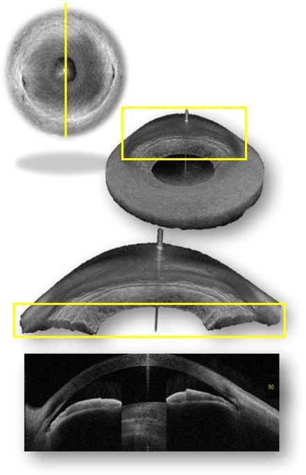
Differentiating the two main forms of primary glaucoma (open-angle and closed-angle glaucoma) depends on the correct assessment of the anterior chamber angle (ACA). This assessment will determine the management plan and prognosis for the disease. The standard method of examining the angle has been, for many years, slit-lamp gonioscopy. This method, although clinically still useful, is less robust for patient follow up and clinical research, given its low reproducibility. Several imaging technologies have been developed in recent years to improve the evaluation of the ACA and overcome the shortcomings of gonioscopy. These recent advances include three-dimensional and 360° analysis by Swept-Source OCT (SS-OCT, CASIA, Tomey, Nagoya, Japan), the introduction of deep learning algorithms for automatic imaging classification and new goniophotographic systems. SS-OCT allows for the first time the assessment of the circumferential extension of angle closure with moderate to good diagnostic performance compared with gonioscopy. Deep learning algorithms are showing promising results for the automation of imaging analysis, and may potentially save physicians’ time in regards of the interpretation of the images. Lastly, goniophotograph systems have the distinct advantage of recordability of gonioscopic findings and are most closely matched to the findings of slit-lamp gonioscopy. 对于主要两种类型的原发性青光眼 (开角型和闭角型青光眼) 的鉴别诊断建立在对前房角进行正确评估的基础上。评估的结果决定青光眼的管理策略和预后。多年来, 检查房角的标准方法一直是裂隙灯前房角镜。尽管该方法目前仍在临床上使用, 但由于其可重复性低, 在病人随访和临床研究中均具有一定缺陷。近年来逐步发展的影像学技术, 不仅能够提高对前房角评估的准确性还能能克服前房角镜检查的缺点。这些新的研究进展包括通过扫频源OCT (SS-OCT, CASIA, Tomey, Nagoya, Japan)进行三维及360°分析, 使用深度学习算法进行图片自动分类, 以及新的房角摄像系统。与前房角镜检查相比, SS-OCT在初次评估房角关闭的范围的评估方面可达中等至良好。深度学习算法展现了令人鼓舞的结果, 这将可能节省医生们分析图像所花费的时间。最后一点, 房角摄像系统最显著的优点是可以记录前房角镜的检查结果, 并且与裂隙灯房角镜的检查结果最匹配。
中文翻译:

前房角成像的最新进展
Differentiating the two main forms of primary glaucoma (open-angle and closed-angle glaucoma) depends on the correct assessment of the anterior chamber angle (ACA). This assessment will determine the management plan and prognosis for the disease. The standard method of examining the angle has been, for many years, slit-lamp gonioscopy. This method, although clinically still useful, is less robust for patient follow up and clinical research, given its low reproducibility. Several imaging technologies have been developed in recent years to improve the evaluation of the ACA and overcome the shortcomings of gonioscopy. These recent advances include three-dimensional and 360° analysis by Swept-Source OCT (SS-OCT, CASIA, Tomey, Nagoya, Japan), the introduction of deep learning algorithms for automatic imaging classification and new goniophotographic systems. SS-OCT allows for the first time the assessment of the circumferential extension of angle closure with moderate to good diagnostic performance compared with gonioscopy. Deep learning algorithms are showing promising results for the automation of imaging analysis, and may potentially save physicians’ time in regards of the interpretation of the images. Lastly, goniophotograph systems have the distinct advantage of recordability of gonioscopic findings and are most closely matched to the findings of slit-lamp gonioscopy. 对于主要两种类型的原发性青光眼 (开角型和闭角型青光眼) 的鉴别诊断建立在对前房角进行正确评估的基础上。评估的结果决定青光眼的管理策略和预后。多年来, 检查房角的标准方法一直是裂隙灯前房角镜。尽管该方法目前仍在临床上使用, 但由于其可重复性低, 在病人随访和临床研究中均具有一定缺陷。近年来逐步发展的影像学技术, 不仅能够提高对前房角评估的准确性还能能克服前房角镜检查的缺点。这些新的研究进展包括通过扫频源OCT (SS-OCT, CASIA, Tomey, Nagoya, Japan)进行三维及360°分析, 使用深度学习算法进行图片自动分类, 以及新的房角摄像系统。与前房角镜检查相比, SS-OCT在初次评估房角关闭的范围的评估方面可达中等至良好。深度学习算法展现了令人鼓舞的结果, 这将可能节省医生们分析图像所花费的时间。最后一点, 房角摄像系统最显著的优点是可以记录前房角镜的检查结果, 并且与裂隙灯房角镜的检查结果最匹配。
更新日期:2019-10-30
中文翻译:

前房角成像的最新进展
Differentiating the two main forms of primary glaucoma (open-angle and closed-angle glaucoma) depends on the correct assessment of the anterior chamber angle (ACA). This assessment will determine the management plan and prognosis for the disease. The standard method of examining the angle has been, for many years, slit-lamp gonioscopy. This method, although clinically still useful, is less robust for patient follow up and clinical research, given its low reproducibility. Several imaging technologies have been developed in recent years to improve the evaluation of the ACA and overcome the shortcomings of gonioscopy. These recent advances include three-dimensional and 360° analysis by Swept-Source OCT (SS-OCT, CASIA, Tomey, Nagoya, Japan), the introduction of deep learning algorithms for automatic imaging classification and new goniophotographic systems. SS-OCT allows for the first time the assessment of the circumferential extension of angle closure with moderate to good diagnostic performance compared with gonioscopy. Deep learning algorithms are showing promising results for the automation of imaging analysis, and may potentially save physicians’ time in regards of the interpretation of the images. Lastly, goniophotograph systems have the distinct advantage of recordability of gonioscopic findings and are most closely matched to the findings of slit-lamp gonioscopy. 对于主要两种类型的原发性青光眼 (开角型和闭角型青光眼) 的鉴别诊断建立在对前房角进行正确评估的基础上。评估的结果决定青光眼的管理策略和预后。多年来, 检查房角的标准方法一直是裂隙灯前房角镜。尽管该方法目前仍在临床上使用, 但由于其可重复性低, 在病人随访和临床研究中均具有一定缺陷。近年来逐步发展的影像学技术, 不仅能够提高对前房角评估的准确性还能能克服前房角镜检查的缺点。这些新的研究进展包括通过扫频源OCT (SS-OCT, CASIA, Tomey, Nagoya, Japan)进行三维及360°分析, 使用深度学习算法进行图片自动分类, 以及新的房角摄像系统。与前房角镜检查相比, SS-OCT在初次评估房角关闭的范围的评估方面可达中等至良好。深度学习算法展现了令人鼓舞的结果, 这将可能节省医生们分析图像所花费的时间。最后一点, 房角摄像系统最显著的优点是可以记录前房角镜的检查结果, 并且与裂隙灯房角镜的检查结果最匹配。


 京公网安备 11010802027423号