当前位置:
X-MOL 学术
›
ACS Appl. Bio Mater.
›
论文详情
Our official English website, www.x-mol.net, welcomes your
feedback! (Note: you will need to create a separate account there.)
nCP:Fe—A Biomineral Magnetic Nanocontrast Agent for Tracking Implanted Stem Cells in Brain Using MRI
ACS Applied Bio Materials ( IF 4.6 ) Pub Date : 2019-10-28 , DOI: 10.1021/acsabm.9b00709
Ida M Anna 1 , Binulal N Sathy 1 , Anusha Ashokan 1 , Genekehal Siddaramana Gowd 1 , Ranjith Ramachandran 1 , Ayalur Kodakara Kochugovindan Unni 2 , Maneesh Manohar 1 , DeepthiMol Chulliyath 1 , Shantikumar Nair 1 , Kishore Bhakoo 3 , Manzoor Koyakutty 1
Affiliation
|
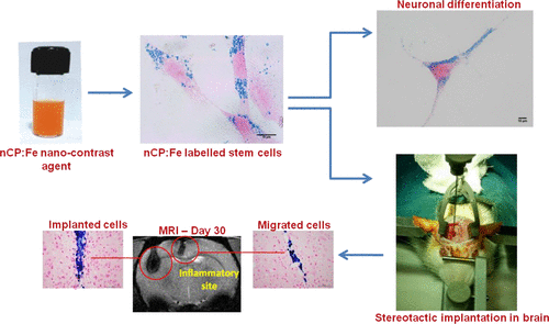
In vivo tracking of transplanted stem cells to monitor their migration, biodistribution, and engraftment in the host tissue is important for assessing the efficacy of stem cell therapeutics. Here, we report a biomineral nanocontrast agent, iron doped calcium phosphate nanoparticles (nCP:Fe), for the in vivo tracking of stem cells in brain using magnetic resonance imaging (MRI). We have synthesized ∼100 nm sized nCP nanoparticles doped with 9.81 wt % Fe3+. In vitro studies using mesenchymal stem cells (MSCs) showed excellent biocompatibility for nCP:Fe with ∼87% labeling efficiency under optimized conditions (100 μg/mL, 6 h). Most importantly, the labeling was not found to affect the neurogenic differentiation potential of MSCs. MRI of labeled cells (∼22.34 pg Fe/cell) showed significant reduction in T2 relaxation time from 195 to 89 ms, rendering dark contrast. In vivo transplantation of labeled cells (1 × 10⁶ cells) in external capsule of healthy rat brain showed a clearly distinguishable hypointense (dark) region in T2 weighted MR images, which remained visible up to 30 days. Subsequently, MRI tracking of labeled MSCs transplanted intracerebrally, 3 mm near to the LPS induced inflammatory site in brain, showed successful migration of labeled MSCs toward the site of inflammation. The cell migration was confirmed ex vivo by Prussian-blue (Fe3+) and Alizarin-red (Ca2+) staining of tissue sections, where individual cells were found migrated to the site of inflammation over a period of 30 days. In summary, our results clearly show that, as a biocompatible mineral composition, nCP:Fe is a promising magnetic nanocontrast agent for MRI based cell tracking in vivo.
中文翻译:

nCP:Fe——一种生物矿物磁性纳米造影剂,用于使用 MRI 跟踪大脑中植入的干细胞
移植干细胞的体内跟踪以监测它们在宿主组织中的迁移、生物分布和植入对于评估干细胞治疗的功效很重要。在这里,我们报告了一种生物矿物纳米造影剂,铁掺杂的磷酸钙纳米粒子 (nCP:Fe),用于使用磁共振成像 (MRI) 对大脑中的干细胞进行体内跟踪。我们合成了掺杂有 9.81 wt% Fe 3+的约 100 nm 尺寸的 nCP 纳米粒子。体外使用间充质干细胞 (MSCs) 的研究表明,在优化条件 (100 μg/mL, 6 h) 下,nCP:Fe 具有优异的生物相容性,标记效率约为 87%。最重要的是,没有发现标记会影响 MSCs 的神经源性分化潜能。标记细胞(~22.34 pg Fe/细胞)的 MRI 显示 T2 弛豫时间从 195 到 89 ms 显着减少,呈现暗对比。标记细胞的体内移植(1 × 10 6健康大鼠脑外囊中的细胞)在 T2 加权 MR 图像中显示出明显可区分的低信号(暗)区域,该区域在 30 天内仍然可见。随后,在脑内 LPS 诱导的炎症部位附近 3 毫米处,对移植到脑内的标记 MSCs 进行 MRI 跟踪,显示标记的 MSCs 成功地向炎症部位迁移。通过对组织切片进行普鲁士蓝 (Fe 3+ ) 和茜素红 (Ca 2+ ) 染色,离体证实了细胞迁移,其中发现单个细胞在 30 天内迁移到炎症部位。总之,我们的研究结果清楚地表明,作为一种生物相容性矿物成分,nCP:Fe 是一种有前途的磁性纳米造影剂,可用于基于 MRI 的细胞追踪体内。
更新日期:2019-10-28
中文翻译:
nCP:Fe——一种生物矿物磁性纳米造影剂,用于使用 MRI 跟踪大脑中植入的干细胞
移植干细胞的体内跟踪以监测它们在宿主组织中的迁移、生物分布和植入对于评估干细胞治疗的功效很重要。在这里,我们报告了一种生物矿物纳米造影剂,铁掺杂的磷酸钙纳米粒子 (nCP:Fe),用于使用磁共振成像 (MRI) 对大脑中的干细胞进行体内跟踪。我们合成了掺杂有 9.81 wt% Fe 3+的约 100 nm 尺寸的 nCP 纳米粒子。体外使用间充质干细胞 (MSCs) 的研究表明,在优化条件 (100 μg/mL, 6 h) 下,nCP:Fe 具有优异的生物相容性,标记效率约为 87%。最重要的是,没有发现标记会影响 MSCs 的神经源性分化潜能。标记细胞(~22.34 pg Fe/细胞)的 MRI 显示 T2 弛豫时间从 195 到 89 ms 显着减少,呈现暗对比。标记细胞的体内移植(1 × 10 6健康大鼠脑外囊中的细胞)在 T2 加权 MR 图像中显示出明显可区分的低信号(暗)区域,该区域在 30 天内仍然可见。随后,在脑内 LPS 诱导的炎症部位附近 3 毫米处,对移植到脑内的标记 MSCs 进行 MRI 跟踪,显示标记的 MSCs 成功地向炎症部位迁移。通过对组织切片进行普鲁士蓝 (Fe 3+ ) 和茜素红 (Ca 2+ ) 染色,离体证实了细胞迁移,其中发现单个细胞在 30 天内迁移到炎症部位。总之,我们的研究结果清楚地表明,作为一种生物相容性矿物成分,nCP:Fe 是一种有前途的磁性纳米造影剂,可用于基于 MRI 的细胞追踪体内。


 京公网安备 11010802027423号