当前位置:
X-MOL 学术
›
Nat. Commun.
›
论文详情
Our official English website, www.x-mol.net, welcomes your
feedback! (Note: you will need to create a separate account there.)
Rapid single-wavelength lightsheet localization microscopy for clarified tissue.
Nature Communications ( IF 14.7 ) Pub Date : 2019-10-18 , DOI: 10.1038/s41467-019-12715-3 Li-An Chu , Chieh-Han Lu , Shun-Min Yang , Yen-Ting Liu , Kuan-Lin Feng , Yun-Chi Tsai , Wei-Kun Chang , Wen-Cheng Wang , Shu-Wei Chang , Peilin Chen , Ting-Kuo Lee , Yeu-Kuang Hwu , Ann-Shyn Chiang , Bi-Chang Chen
Nature Communications ( IF 14.7 ) Pub Date : 2019-10-18 , DOI: 10.1038/s41467-019-12715-3 Li-An Chu , Chieh-Han Lu , Shun-Min Yang , Yen-Ting Liu , Kuan-Lin Feng , Yun-Chi Tsai , Wei-Kun Chang , Wen-Cheng Wang , Shu-Wei Chang , Peilin Chen , Ting-Kuo Lee , Yeu-Kuang Hwu , Ann-Shyn Chiang , Bi-Chang Chen

|
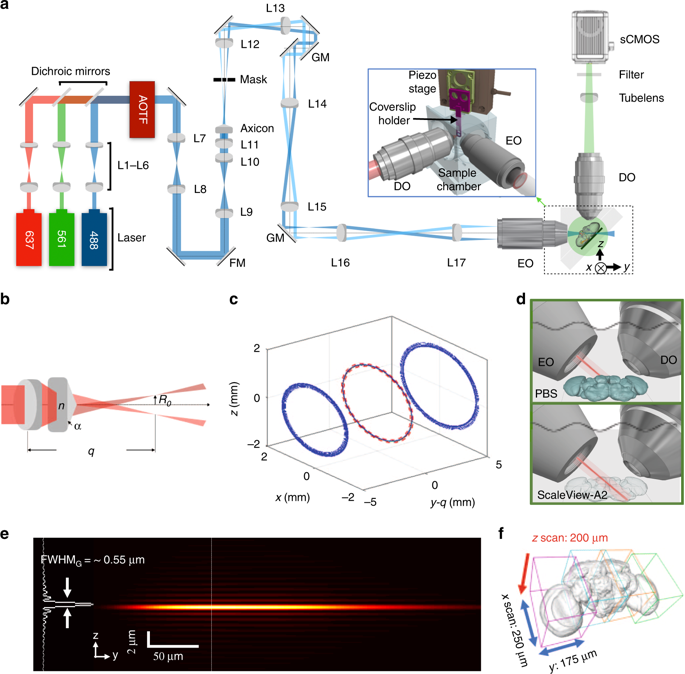
Optical super-resolution microscopy allows nanoscale imaging of protein molecules in intact biological tissues. However, it is still challenging to perform large volume super-resolution imaging for entire animal organs. Here we develop a single-wavelength Bessel lightsheet method, optimized for refractive-index matching with clarified specimens to overcome the aberrations encountered in imaging thick tissues. Using spontaneous blinking fluorophores to label proteins of interest, we resolve the morphology of most, if not all, dopaminergic neurons in the whole adult brain (3.64 × 107 µm3) of Drosophila melanogaster at the nanometer scale with high imaging speed (436 µm3 per second) for localization. Quantitative single-molecule localization reveals the subcellular distribution of a monoamine transporter protein in the axons of a single, identified serotonergic Dorsal Paired Medial (DPM) neuron. Large datasets are obtained from imaging one brain per day to provide a robust statistical analysis of these imaging data.
中文翻译:

快速单波长光片定位显微镜用于澄清的组织。
光学超分辨率显微镜可以对完整的生物组织中的蛋白质分子进行纳米级成像。然而,对整个动物器官进行大体积超分辨率成像仍然是一项挑战。在这里,我们开发了一种单波长贝塞尔(Bessel)光片法,该方法针对与澄清标本的折射率匹配进行了优化,以克服在对厚组织成像时遇到的像差。使用自发闪烁的荧光团标记感兴趣的蛋白质,我们以纳米级的速度以每秒436 µm3的纳米级分辨率分辨了果蝇整个成年大脑(3.64×107 µm3)中大多数(如果不是全部)多巴胺能神经元的形态。 )进行本地化。定量单分子定位揭示了单胺转运蛋白在单个轴突中的亚细胞分布,鉴定出血清素能神经对配对内侧神经元(DPM)。每天从一个大脑成像获得大量数据集,以提供对这些成像数据的可靠统计分析。
更新日期:2019-10-19
中文翻译:

快速单波长光片定位显微镜用于澄清的组织。
光学超分辨率显微镜可以对完整的生物组织中的蛋白质分子进行纳米级成像。然而,对整个动物器官进行大体积超分辨率成像仍然是一项挑战。在这里,我们开发了一种单波长贝塞尔(Bessel)光片法,该方法针对与澄清标本的折射率匹配进行了优化,以克服在对厚组织成像时遇到的像差。使用自发闪烁的荧光团标记感兴趣的蛋白质,我们以纳米级的速度以每秒436 µm3的纳米级分辨率分辨了果蝇整个成年大脑(3.64×107 µm3)中大多数(如果不是全部)多巴胺能神经元的形态。 )进行本地化。定量单分子定位揭示了单胺转运蛋白在单个轴突中的亚细胞分布,鉴定出血清素能神经对配对内侧神经元(DPM)。每天从一个大脑成像获得大量数据集,以提供对这些成像数据的可靠统计分析。


 京公网安备 11010802027423号