当前位置:
X-MOL 学术
›
Acta Pharm. Sin. B
›
论文详情
Our official English website, www.x-mol.net, welcomes your feedback! (Note: you will need to create a separate account there.)
Novel radioligands for imaging sigma-1 receptor in brain using positron emission tomography (PET).
Acta Pharmaceutica Sinica B ( IF 14.7 ) Pub Date : 2019-07-11 , DOI: 10.1016/j.apsb.2019.07.002 Yu Lan 1, 2 , Ping Bai 1 , Zude Chen 1 , Ramesh Neelamegam 3 , Michael S Placzek 1 , Hao Wang 1 , Stephanie A Fiedler 1 , Jing Yang 1 , Gengyang Yuan 3 , Xiying Qu 3 , Hayden R Schmidt 4 , Jinchun Song 2 , Marc D Normandin 3 , Chongzhao Ran 1 , Changning Wang 1
Acta Pharmaceutica Sinica B ( IF 14.7 ) Pub Date : 2019-07-11 , DOI: 10.1016/j.apsb.2019.07.002 Yu Lan 1, 2 , Ping Bai 1 , Zude Chen 1 , Ramesh Neelamegam 3 , Michael S Placzek 1 , Hao Wang 1 , Stephanie A Fiedler 1 , Jing Yang 1 , Gengyang Yuan 3 , Xiying Qu 3 , Hayden R Schmidt 4 , Jinchun Song 2 , Marc D Normandin 3 , Chongzhao Ran 1 , Changning Wang 1
Affiliation
|
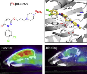
The sigma-1 receptor (σ 1R) is a unique intracellular protein. σ 1R plays a major role in various pathological conditions in the central nervous system (CNS), implicated in several neuropsychiatric disorders. Imaging of σ 1R in the brain using positron emission tomography (PET) could serve as a noninvasively tool for enhancing the understanding of the disease's pathophysiology. Moreover, σ 1R PET tracers can be used for target validation and quantification in diagnosis. Herein, we describe the radiosynthesis, in vivo PET/CT imaging of novel σ 1R 11C-labeled radioligands based on 6-hydroxypyridazinone, [11C]HCC0923 and [11C]HCC0929. Two radioligands have high affinities to σ 1R, with good selectivity. In mice PET/CT imaging, both radioligands showed appropriate kinetics and distributions. Additionally, the specific interactions of two radioligands were reduced by compounds 13 and 15 (self-blocking). Of the two, [11C]HCC0929 was further investigated in positive ligands blocking studies, using classic σ 1R agonist SA 4503 and σ 1R antagonist PD 144418. Both σ 1R ligands could extensively decreased the uptake of [11C]HCC0929 in mice brain. Besides, the biodistribution of major brain regions and organs of mice were determined in vivo. These studies demonstrated that two radioligands, especially [11C]HCC0929, possessed ideal imaging properties and might be valuable tools for non-invasive quantification of σ 1R in brain.
中文翻译:

使用正电子发射断层扫描(PET)成像大脑中sigma-1受体的新型放射性配体。
sigma-1受体(σ1R)是一种独特的细胞内蛋白。σ1R在中枢神经系统(CNS)的各种病理状况中起主要作用,与多种神经精神疾病有关。使用正电子发射断层扫描(PET)在大脑中对σ1R进行成像可作为一种非侵入性工具,用于增强对该疾病的病理生理学的理解。此外,σ1R PET示踪剂可用于目标验证和诊断定量。在这里,我们描述了基于6-羟基哒嗪酮,[11C] HCC0923和[11C] HCC0929的新型σ1R 11C标记的放射性配体的放射合成,体内PET / CT成像。两个放射性配体对σ1R具有高亲和力,并且具有良好的选择性。在小鼠PET / CT成像中,两种放射性配体均显示出适当的动力学和分布。此外,化合物13和15降低了两个放射性配体的特异性相互作用(自封闭)。在这两种化合物中,[11C] HCC0929在经典的σ1R激动剂SA 4503和σ1R拮抗剂PD 144418的阳性配体阻断研究中作了进一步研究。这两个σ1R配体均可广泛降低小鼠脑中对[11C] HCC0929的吸收。此外,体内主要小鼠的大脑区域和器官的生物分布被确定。这些研究表明,两个放射性配体,特别是[11C] HCC0929,具有理想的成像特性,可能是对脑中σ1R进行无创定量的有价值的工具。两种σ1R配体均可广泛降低小鼠脑中对[11C] HCC0929的吸收。此外,体内主要小鼠的大脑区域和器官的生物分布被确定。这些研究表明,两个放射性配体,特别是[11C] HCC0929,具有理想的成像特性,可能是对脑中σ1R进行无创定量的有价值的工具。两种σ1R配体均可广泛降低小鼠脑中对[11C] HCC0929的吸收。此外,体内主要小鼠的大脑区域和器官的生物分布被确定。这些研究表明,两个放射性配体,特别是[11C] HCC0929,具有理想的成像特性,可能是对脑中σ1R进行无创定量的有价值的工具。
更新日期:2019-09-09
中文翻译:
使用正电子发射断层扫描(PET)成像大脑中sigma-1受体的新型放射性配体。
sigma-1受体(σ1R)是一种独特的细胞内蛋白。σ1R在中枢神经系统(CNS)的各种病理状况中起主要作用,与多种神经精神疾病有关。使用正电子发射断层扫描(PET)在大脑中对σ1R进行成像可作为一种非侵入性工具,用于增强对该疾病的病理生理学的理解。此外,σ1R PET示踪剂可用于目标验证和诊断定量。在这里,我们描述了基于6-羟基哒嗪酮,[11C] HCC0923和[11C] HCC0929的新型σ1R 11C标记的放射性配体的放射合成,体内PET / CT成像。两个放射性配体对σ1R具有高亲和力,并且具有良好的选择性。在小鼠PET / CT成像中,两种放射性配体均显示出适当的动力学和分布。此外,化合物13和15降低了两个放射性配体的特异性相互作用(自封闭)。在这两种化合物中,[11C] HCC0929在经典的σ1R激动剂SA 4503和σ1R拮抗剂PD 144418的阳性配体阻断研究中作了进一步研究。这两个σ1R配体均可广泛降低小鼠脑中对[11C] HCC0929的吸收。此外,体内主要小鼠的大脑区域和器官的生物分布被确定。这些研究表明,两个放射性配体,特别是[11C] HCC0929,具有理想的成像特性,可能是对脑中σ1R进行无创定量的有价值的工具。两种σ1R配体均可广泛降低小鼠脑中对[11C] HCC0929的吸收。此外,体内主要小鼠的大脑区域和器官的生物分布被确定。这些研究表明,两个放射性配体,特别是[11C] HCC0929,具有理想的成像特性,可能是对脑中σ1R进行无创定量的有价值的工具。两种σ1R配体均可广泛降低小鼠脑中对[11C] HCC0929的吸收。此外,体内主要小鼠的大脑区域和器官的生物分布被确定。这些研究表明,两个放射性配体,特别是[11C] HCC0929,具有理想的成像特性,可能是对脑中σ1R进行无创定量的有价值的工具。


 京公网安备 11010802027423号