当前位置:
X-MOL 学术
›
J. Biophotonics
›
论文详情
Our official English website, www.x-mol.net, welcomes your
feedback! (Note: you will need to create a separate account there.)
Neurosurgical brain tumor detection based on intraoperative optical intrinsic signal imaging technique: A case report of glioblastoma.
Journal of Biophotonics ( IF 2.0 ) Pub Date : 2019-10-29 , DOI: 10.1002/jbio.201900200 Xin-Rui Liu,Tien-Yu Hsiao,Yun-Qian Li,Kai-Shih Chiu,Chun-Jung Huang,Shan-Ji Li,Ching Po Lin,Gang Zhao,Chia-Wei Sun
Journal of Biophotonics ( IF 2.0 ) Pub Date : 2019-10-29 , DOI: 10.1002/jbio.201900200 Xin-Rui Liu,Tien-Yu Hsiao,Yun-Qian Li,Kai-Shih Chiu,Chun-Jung Huang,Shan-Ji Li,Ching Po Lin,Gang Zhao,Chia-Wei Sun

|
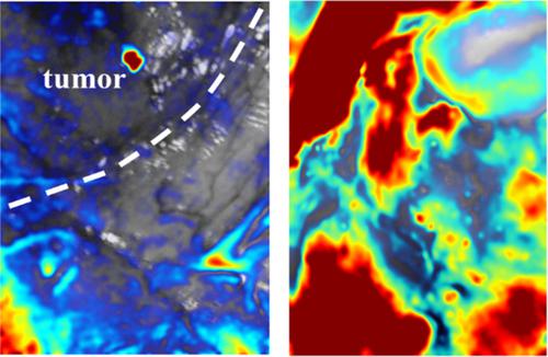
The delineation of brain tumor margins has been a challenging objective in neurosurgery for decades. Despite the development of various preoperative imaging techniques, the current methodology is still insufficient for clinical practice. We present an intraoperative optical intrinsic signal imaging system for brain tumor surgery and establish a data processing procedure model to localize tumors. From the experimental result of a glioblastoma patient, we observe a relative small oscillation of ΔHbD in tumor region and speculate that vessels in tumor region have poor ability to provide oxygen. We applied the same data processing procedure on the second time data and proclaimed a successful surgery. Figure: Merged ΔHbD image captured prior and posterior to tumor removal.
中文翻译:

基于术中光学内在信号成像技术的神经外科脑瘤检测:胶质母细胞瘤的一例报道。
数十年来,脑肿瘤边缘的描绘一直是神经外科手术中的一个具有挑战性的目标。尽管各种术前成像技术得到了发展,但是当前的方法仍不足以用于临床实践。我们提出了一种用于脑肿瘤手术的术中光学内在信号成像系统,并建立了用于定位肿瘤的数据处理程序模型。从胶质母细胞瘤患者的实验结果中,我们观察到肿瘤区域中ΔHbD的振荡相对较小,并推测肿瘤区域中的血管提供氧气的能力较弱。我们对第二次数据应用了相同的数据处理程序,并宣布手术成功。图:在切除肿瘤之前和之后捕获的合并的ΔHbD图像。
更新日期:2019-10-29
中文翻译:

基于术中光学内在信号成像技术的神经外科脑瘤检测:胶质母细胞瘤的一例报道。
数十年来,脑肿瘤边缘的描绘一直是神经外科手术中的一个具有挑战性的目标。尽管各种术前成像技术得到了发展,但是当前的方法仍不足以用于临床实践。我们提出了一种用于脑肿瘤手术的术中光学内在信号成像系统,并建立了用于定位肿瘤的数据处理程序模型。从胶质母细胞瘤患者的实验结果中,我们观察到肿瘤区域中ΔHbD的振荡相对较小,并推测肿瘤区域中的血管提供氧气的能力较弱。我们对第二次数据应用了相同的数据处理程序,并宣布手术成功。图:在切除肿瘤之前和之后捕获的合并的ΔHbD图像。


 京公网安备 11010802027423号