Our official English website, www.x-mol.net, welcomes your
feedback! (Note: you will need to create a separate account there.)
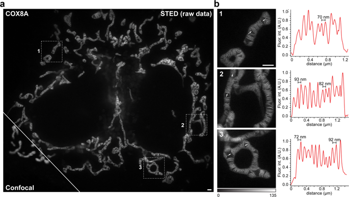
Live-cell STED nanoscopy of mitochondrial cristae.
Scientific Reports ( IF 3.8 ) Pub Date : 2019-08-27 , DOI: 10.1038/s41598-019-48838-2 Till Stephan 1, 2 , Axel Roesch 1, 2 , Dietmar Riedel 3 , Stefan Jakobs 1, 2
Scientific Reports ( IF 3.8 ) Pub Date : 2019-08-27 , DOI: 10.1038/s41598-019-48838-2 Till Stephan 1, 2 , Axel Roesch 1, 2 , Dietmar Riedel 3 , Stefan Jakobs 1, 2
Affiliation
|
Mitochondria are highly dynamic organelles that exhibit a complex inner architecture. They exhibit a smooth outer membrane and a highly convoluted inner membrane that forms invaginations called cristae. Imaging cristae in living cells poses a formidable challenge for super-resolution light microscopy. Relying on a cell line stably expressing the mitochondrial protein COX8A fused to the SNAP-tag and using STED (stimulated emission depletion) nanoscopy, we demonstrate the visualization of cristae dynamics in cultivated human cells. We show that in human HeLa cells lamellar cristae are often arranged in groups separated by voids that are generally occupied by mitochondrial nucleoids.
中文翻译:

线粒体cr的活细胞STED纳米显微镜检查。
线粒体是高度动态的细胞器,具有复杂的内部结构。它们表现出光滑的外膜和高度卷曲的内膜,形成称为cr的内陷。活细胞中的cr缝成像对超分辨率光学显微镜提出了巨大的挑战。依靠稳定表达融合到SNAP标签的线粒体蛋白COX8A的细胞系,并使用STED(受激发射损耗)纳米显微镜,我们证明了培养的人类细胞中ista动力学的可视化。我们显示,在人类HeLa细胞中,层状cr通常以通常由线粒体核苷酸占据的空隙分隔成组。
更新日期:2019-08-27
中文翻译:
线粒体cr的活细胞STED纳米显微镜检查。
线粒体是高度动态的细胞器,具有复杂的内部结构。它们表现出光滑的外膜和高度卷曲的内膜,形成称为cr的内陷。活细胞中的cr缝成像对超分辨率光学显微镜提出了巨大的挑战。依靠稳定表达融合到SNAP标签的线粒体蛋白COX8A的细胞系,并使用STED(受激发射损耗)纳米显微镜,我们证明了培养的人类细胞中ista动力学的可视化。我们显示,在人类HeLa细胞中,层状cr通常以通常由线粒体核苷酸占据的空隙分隔成组。


 京公网安备 11010802027423号