当前位置:
X-MOL 学术
›
Light Sci. Appl.
›
论文详情
Our official English website, www.x-mol.net, welcomes your
feedback! (Note: you will need to create a separate account there.)
Multiplexed laser particles for spatially resolved single-cell analysis.
Light: Science & Applications ( IF 20.6 ) Pub Date : 2019-08-21 , DOI: 10.1038/s41377-019-0183-5 Sheldon J J Kwok 1, 2, 3 , Nicola Martino 1 , Paul H Dannenberg 1, 2 , Seok-Hyun Yun 1, 2
Light: Science & Applications ( IF 20.6 ) Pub Date : 2019-08-21 , DOI: 10.1038/s41377-019-0183-5 Sheldon J J Kwok 1, 2, 3 , Nicola Martino 1 , Paul H Dannenberg 1, 2 , Seok-Hyun Yun 1, 2
Affiliation
|
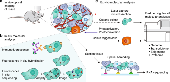
Biomolecular analysis at the single-cell level is increasingly important in the study of cellular heterogeneity and its consequences, particularly in organismic development and complex diseases such as cancer. Single-cell molecular analyses have led to the identification of new cell types1 and the discovery of novel targets for diagnosis and therapy2. While these analyses are performed predominantly on dissociated single cells, emerging techniques seek understanding of cellular state, cellular function and cell-cell interactions within the native tissue environment by combining optical microscopy and single-cell molecular analyses. These techniques include in situ multiplexed imaging of fluorescently labeled proteins and nucleotides, as well as low-throughput ex vivo methods in which specific cells are isolated for downstream molecular analyses. However, these methods are limited in either the number and type of molecular species they can identify or the number of cells that can be analyzed. High-throughput methods are needed for comprehensive profiling of many cells (>1000) to detect rare cell types, discriminate relevant biomarkers from intrinsic population noise, and reduce the time and cost of measurement. Many established, high-throughput single-cell analyses are not directly applicable because they require tissue dissociation, leading to a loss of spatial information3. No current methods exist that can seamlessly connect spatial mapping to single-cell techniques. In this Perspective, we review current methods for spatially resolved single-cell analysis and discuss the prospect of novel multiplexed imaging probes, called laser particles, which allow individual cells to be tagged in tissue and analyzed subsequently using high-throughput, comprehensive single-cell techniques.
中文翻译:

用于空间分辨单细胞分析的多重激光粒子。
单细胞水平的生物分子分析在细胞异质性及其后果的研究中变得越来越重要,特别是在有机体发育和癌症等复杂疾病中。单细胞分子分析有助于识别新的细胞类型1并发现新的诊断和治疗靶标2。虽然这些分析主要针对分离的单细胞进行,但新兴技术通过结合光学显微镜和单细胞分子分析来寻求对天然组织环境中的细胞状态、细胞功能和细胞间相互作用的理解。这些技术包括荧光标记的蛋白质和核苷酸的原位多重成像,以及分离特定细胞进行下游分子分析的低通量离体方法。然而,这些方法在它们可以识别的分子种类的数量和类型或可以分析的细胞数量方面受到限制。需要高通量方法对许多细胞(> 1000)进行全面分析,以检测稀有细胞类型,将相关生物标志物与固有群体噪声区分开来,并减少测量时间和成本。许多已建立的高通量单细胞分析并不直接适用,因为它们需要组织解离,从而导致空间信息丢失3。目前尚不存在能够将空间映射与单细胞技术无缝连接的方法。在本视角中,我们回顾了当前的空间分辨单细胞分析方法,并讨论了新型多重成像探针(称为激光粒子)的前景,该探针允许在组织中标记单个细胞,并随后使用高通量、全面的单细胞进行分析技术。
更新日期:2019-08-20
中文翻译:
用于空间分辨单细胞分析的多重激光粒子。
单细胞水平的生物分子分析在细胞异质性及其后果的研究中变得越来越重要,特别是在有机体发育和癌症等复杂疾病中。单细胞分子分析有助于识别新的细胞类型1并发现新的诊断和治疗靶标2。虽然这些分析主要针对分离的单细胞进行,但新兴技术通过结合光学显微镜和单细胞分子分析来寻求对天然组织环境中的细胞状态、细胞功能和细胞间相互作用的理解。这些技术包括荧光标记的蛋白质和核苷酸的原位多重成像,以及分离特定细胞进行下游分子分析的低通量离体方法。然而,这些方法在它们可以识别的分子种类的数量和类型或可以分析的细胞数量方面受到限制。需要高通量方法对许多细胞(> 1000)进行全面分析,以检测稀有细胞类型,将相关生物标志物与固有群体噪声区分开来,并减少测量时间和成本。许多已建立的高通量单细胞分析并不直接适用,因为它们需要组织解离,从而导致空间信息丢失3。目前尚不存在能够将空间映射与单细胞技术无缝连接的方法。在本视角中,我们回顾了当前的空间分辨单细胞分析方法,并讨论了新型多重成像探针(称为激光粒子)的前景,该探针允许在组织中标记单个细胞,并随后使用高通量、全面的单细胞进行分析技术。


 京公网安备 11010802027423号