当前位置:
X-MOL 学术
›
J. Control. Release
›
论文详情
Our official English website, www.x-mol.net, welcomes your
feedback! (Note: you will need to create a separate account there.)
Integrin α6 targeted positron emission tomography imaging of hepatocellular carcinoma in mouse models.
Journal of Controlled Release ( IF 10.5 ) Pub Date : 2019-08-07 , DOI: 10.1016/j.jconrel.2019.08.003 Guo-Kai Feng 1 , Jia-Cong Ye 1 , Wei-Guang Zhang 1 , Yan Mei 1 , Chao Zhou 1 , Yi-Tai Xiao 1 , Xin-Ling Li 1 , Wei Fan 1 , Fan Wang 2 , Mu-Sheng Zeng 1
Journal of Controlled Release ( IF 10.5 ) Pub Date : 2019-08-07 , DOI: 10.1016/j.jconrel.2019.08.003 Guo-Kai Feng 1 , Jia-Cong Ye 1 , Wei-Guang Zhang 1 , Yan Mei 1 , Chao Zhou 1 , Yi-Tai Xiao 1 , Xin-Ling Li 1 , Wei Fan 1 , Fan Wang 2 , Mu-Sheng Zeng 1
Affiliation

|
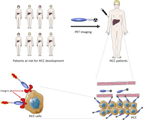
Integrin α6 emerges to be a diagnostic biomarker for hepatocellular carcinoma (HCC). Here, we translated our previously identified integrin α6 targeted peptide RWY into a positron emission tomography (PET) tracer 18F-RWY for the detection of HCC lesions in following four HCC mouse models including subcutaneous, orthotopic, genetically engineered and chemical induced HCC mice. 18F-RWY produced high PET signals in liver tumor tissues that were reduced by blocking studies using nonradiolabeled RWY peptide. We compared the integrin α6 targeted PET tracer 18F-RWY with the integrin αvβ3-targeted PET tracer 18F-3PRGD2 and the clinical PET tracer 18F-FDG in chemical induced HCC mice. Among 12 HCC identified by enhanced magnetic resonance imaging (MRI) with hepatocellular specific gadoxetate disodium Gd-EOB-DTPA, the sensitivities of 18F-RWY, 18F-3PRGD2 and 18F-FDG were approximately 92%, 73% and 50% while the tumor-to-liver ratios were 4.36 ± 1.41, 1.97 ± 0.43 and 1.63 ± 0.23 respectively. Additionally, PET imaging with the integrin α6 targeted 18F-RWY enabled to visualize small HCC lesions with diameters approximately 0.2 cm that was hard to be distinguished from surround hepatic vascular by enhanced MRI with Gd-EOB-DTPA. These findings potentiate the use of integrin α6 targeted PET tracer 18F-RWY for the detection of HCC.
中文翻译:

整合素α6靶向肝细胞癌小鼠模型中的正电子发射断层显像。
整联蛋白α6成为肝细胞癌(HCC)的诊断生物标志物。在这里,我们将先前鉴定的整合素α6靶向肽RWY转换为正电子发射断层扫描(PET)示踪剂18F-RWY,用于在以下四种HCC小鼠模型中检测HCC病变,包括皮下,原位,基因工程和化学诱导的HCC小鼠。18F-RWY在肝肿瘤组织中产生高PET信号,但通过使用非放射性标记的RWY肽的阻断研究降低了18F-RWY的信号。我们在化学诱导的HCC小鼠中比较了针对整合素α6的PET示踪剂18F-RWY与针对整合素αvβ3的PET示踪剂18F-3PRGD2和临床PET示踪剂18F-FDG。在通过增强磁共振成像(MRI)鉴定的12种肝细胞癌中,肝细胞特异的gadoxetate二钠Gd-EOB-DTPA有18F-RWY的敏感性,18F-3PRGD2和18F-FDG分别约为92%,73%和50%,而肿瘤与肝脏的比率分别为4.36±1.41、1.97±0.43和1.63±0.23。此外,以整联蛋白α6为靶标的18F-RWY进行的PET成像能够可视化直径约为0.2 cm的小HCC病变,通过Gd-EOB-DTPA增强的MRI很难将其与周围的肝血管区分开。这些发现增强了将整联蛋白α6靶向的PET示踪剂18F-RWY用于检测HCC的能力。用Gd-EOB-DTPA增强的MRI很难将其与周围的肝血管区分开2 cm。这些发现增强了将整联蛋白α6靶向的PET示踪剂18F-RWY用于检测HCC的能力。用Gd-EOB-DTPA增强的MRI很难将其与周围的肝血管区分开2 cm。这些发现增强了将整联蛋白α6靶向的PET示踪剂18F-RWY用于检测HCC的能力。
更新日期:2019-08-07
中文翻译:

整合素α6靶向肝细胞癌小鼠模型中的正电子发射断层显像。
整联蛋白α6成为肝细胞癌(HCC)的诊断生物标志物。在这里,我们将先前鉴定的整合素α6靶向肽RWY转换为正电子发射断层扫描(PET)示踪剂18F-RWY,用于在以下四种HCC小鼠模型中检测HCC病变,包括皮下,原位,基因工程和化学诱导的HCC小鼠。18F-RWY在肝肿瘤组织中产生高PET信号,但通过使用非放射性标记的RWY肽的阻断研究降低了18F-RWY的信号。我们在化学诱导的HCC小鼠中比较了针对整合素α6的PET示踪剂18F-RWY与针对整合素αvβ3的PET示踪剂18F-3PRGD2和临床PET示踪剂18F-FDG。在通过增强磁共振成像(MRI)鉴定的12种肝细胞癌中,肝细胞特异的gadoxetate二钠Gd-EOB-DTPA有18F-RWY的敏感性,18F-3PRGD2和18F-FDG分别约为92%,73%和50%,而肿瘤与肝脏的比率分别为4.36±1.41、1.97±0.43和1.63±0.23。此外,以整联蛋白α6为靶标的18F-RWY进行的PET成像能够可视化直径约为0.2 cm的小HCC病变,通过Gd-EOB-DTPA增强的MRI很难将其与周围的肝血管区分开。这些发现增强了将整联蛋白α6靶向的PET示踪剂18F-RWY用于检测HCC的能力。用Gd-EOB-DTPA增强的MRI很难将其与周围的肝血管区分开2 cm。这些发现增强了将整联蛋白α6靶向的PET示踪剂18F-RWY用于检测HCC的能力。用Gd-EOB-DTPA增强的MRI很难将其与周围的肝血管区分开2 cm。这些发现增强了将整联蛋白α6靶向的PET示踪剂18F-RWY用于检测HCC的能力。


 京公网安备 11010802027423号