Our official English website, www.x-mol.net, welcomes your
feedback! (Note: you will need to create a separate account there.)
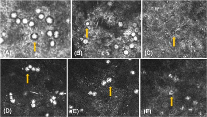
Clinical features and serial changes of Acanthamoeba keratitis: an in vivo confocal microscopy study
Eye ( IF 2.8 ) Pub Date : 2019-07-10 , DOI: 10.1038/s41433-019-0482-3 Suxia Li 1, 2 , Jiang Bian 2 , Yuting Wang 2 , Shuting Wang 2 , Xin Wang 2 , Weiyun Shi 1, 2
Eye ( IF 2.8 ) Pub Date : 2019-07-10 , DOI: 10.1038/s41433-019-0482-3 Suxia Li 1, 2 , Jiang Bian 2 , Yuting Wang 2 , Shuting Wang 2 , Xin Wang 2 , Weiyun Shi 1, 2
Affiliation
|
Purpose To observe the clinical features and serial changes of Acanthamoeba keratitis (AK) during medical treatment by using confocal microscopy. Methods Thirty-seven patients (37 eyes) diagnosed with AK were included in this study. Confocal microscopy was used to observe the morphology, distribution, and density of Acanthamoeba cysts before and after medication. The differences between cysts and inflammatory cells were identified. Results Acanthamoeba cysts were detected at a rate of 94.6% (35/37) by repeated confocal microscopic examinations. The cysts consisting of a lowly light-reflective wall and a high-refractive nucleus, showed cluster or chain distribution in the corneal stroma, which was different from inflammatory cells. After medical therapy, the nucleus of cysts or peripheral corneal tissue gradually dissolved to a hollow configuration. Some of the hollow cysts existed for up to 6 months. The quantity of cysts increased after 1–2 weeks of medication in 23 patients (62.1%), and then began to decrease in 13 patients (35.1 %) who were responsive to anti-amoebic treatment. Conclusion Acanthamoeba cysts have many typical clinical features that can be identified by confocal microscopy, which may serve as a valuable tool to guide clinical evaluation and treatment of AK.
中文翻译:

棘阿米巴角膜炎的临床特征和系列变化:体内共聚焦显微镜研究
目的采用共聚焦显微镜观察棘阿米巴角膜炎(AK)在药物治疗过程中的临床特点及系列变化。方法 37 例 37 眼 AK 患者纳入本研究。采用共聚焦显微镜观察用药前后棘阿米巴包囊的形态、分布和密度。确定了囊肿和炎性细胞之间的差异。结果 反复共聚焦显微镜检出棘阿米巴囊肿的检出率为94.6%(35/37)。囊肿由低反光壁和高屈光核组成,在角膜基质中呈簇状或链状分布,与炎症细胞不同。药物治疗后,囊核或周边角膜组织逐渐溶解成空心结构。一些空心囊肿存在长达 6 个月。23 名患者(62.1%)在用药 1-2 周后囊肿数量增加,然后在对抗阿米巴治疗有反应的 13 名患者(35.1%)中开始减少。结论 棘阿米巴囊肿具有许多典型的临床特征,可通过共聚焦显微镜识别,可作为指导AK临床评估和治疗的有价值的工具。
更新日期:2019-07-10
中文翻译:
棘阿米巴角膜炎的临床特征和系列变化:体内共聚焦显微镜研究
目的采用共聚焦显微镜观察棘阿米巴角膜炎(AK)在药物治疗过程中的临床特点及系列变化。方法 37 例 37 眼 AK 患者纳入本研究。采用共聚焦显微镜观察用药前后棘阿米巴包囊的形态、分布和密度。确定了囊肿和炎性细胞之间的差异。结果 反复共聚焦显微镜检出棘阿米巴囊肿的检出率为94.6%(35/37)。囊肿由低反光壁和高屈光核组成,在角膜基质中呈簇状或链状分布,与炎症细胞不同。药物治疗后,囊核或周边角膜组织逐渐溶解成空心结构。一些空心囊肿存在长达 6 个月。23 名患者(62.1%)在用药 1-2 周后囊肿数量增加,然后在对抗阿米巴治疗有反应的 13 名患者(35.1%)中开始减少。结论 棘阿米巴囊肿具有许多典型的临床特征,可通过共聚焦显微镜识别,可作为指导AK临床评估和治疗的有价值的工具。


 京公网安备 11010802027423号