当前位置:
X-MOL 学术
›
ACS Biomater. Sci. Eng.
›
论文详情
Our official English website, www.x-mol.net, welcomes your
feedback! (Note: you will need to create a separate account there.)
Assessment of Enhanced Thermal Effect Due to Gold Nanoparticles during MR-Guided High-Intensity Focused Ultrasound (HIFU) Procedures Using a Mouse-Tumor Model
ACS Biomaterials Science & Engineering ( IF 5.4 ) Pub Date : 2019-06-18 00:00:00 , DOI: 10.1021/acsbiomaterials.9b00368 Surendra B. Devarakonda 1 , Keith Stringer 2 , Marepalli Rao 3 , Matthew Myers 4 , Rupak Banerjee 1
ACS Biomaterials Science & Engineering ( IF 5.4 ) Pub Date : 2019-06-18 00:00:00 , DOI: 10.1021/acsbiomaterials.9b00368 Surendra B. Devarakonda 1 , Keith Stringer 2 , Marepalli Rao 3 , Matthew Myers 4 , Rupak Banerjee 1
Affiliation

|
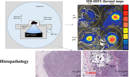
An in vivo study was conducted using a mouse tumor model, to assess the utility of using gold nanoparticles (gNPs) during HIFU procedures to locally enhance heating at low powers. Tumors were grown using melanoma tumor cells (B16/F10) subcutaneously on the right flanks of mice (C57Bl/6J). Physiologically relevant concentrations (0 and 0.125%) of gNPs were directly injected into the tumors. Sonications at acoustic powers of 10 and 30 W were performed for a duration of 16 s inside a magnetic-resonance system. Temperature increases and lesion volumes were calculated and compared for procedures with and without gNPs. Histopathology study was conducted using a cleaved caspase 3 antibody and hematoxylin and eosin staining after removing the tumors from the mice. For an acoustic power of 30 W, end-of-sonication temperature increases of 25.4 ± 3.8 °C (0% gNP) and 42.2 ± 4.6 °C (0.125% gNP) were measured. Using cleaved caspase 3 antibody, it was observed that more than 1% of nuclei are affected in the case of 0.125% and 30 W but only 0.01% of nuclei are affected in the 0% case. For 30 W and a gNP concentration of 0.125%, a lesion volume of 0.33 ± 0.22 mm3 was obtained, while no lesion was observed without gNP’s.
中文翻译:

使用小鼠肿瘤模型评估MR引导的高强度聚焦超声(HIFU)手术过程中由于金纳米粒子引起的增强的热效应
使用小鼠肿瘤模型进行了一项体内研究,以评估在HIFU程序中使用金纳米颗粒(gNP)局部增强低功率加热的效用。使用黑素瘤肿瘤细胞(B16 / F10)在小鼠右腹侧(C57Bl / 6J)皮下生长肿瘤。将生理学上相关浓度(0和0.125%)的gNP直接注入肿瘤中。在磁共振系统内以10瓦和30瓦的声功率进行超声,持续16 s。计算了温度升高和病变体积,并比较了有和没有gNP的程序。从小鼠中取出肿瘤后,使用裂解的caspase 3抗体以及苏木精和曙红染色进行组织病理学研究。对于30 W的声功率,超声处理结束时的温度升高25.4±3。测量了8°C(0%gNP)和42.2±4.6°C(0.125%gNP)。使用裂解的胱天蛋白酶3抗体,观察到在0.125%和30W的情况下,超过1%的细胞核受到影响,而在0%的情况下,仅0.01%的细胞核受到影响。对于30 W和gNP浓度为0.125%的情况,病变体积为0.33±0.22 mm获得3个,而没有gNP则没有观察到病变。
更新日期:2019-06-18
中文翻译:

使用小鼠肿瘤模型评估MR引导的高强度聚焦超声(HIFU)手术过程中由于金纳米粒子引起的增强的热效应
使用小鼠肿瘤模型进行了一项体内研究,以评估在HIFU程序中使用金纳米颗粒(gNP)局部增强低功率加热的效用。使用黑素瘤肿瘤细胞(B16 / F10)在小鼠右腹侧(C57Bl / 6J)皮下生长肿瘤。将生理学上相关浓度(0和0.125%)的gNP直接注入肿瘤中。在磁共振系统内以10瓦和30瓦的声功率进行超声,持续16 s。计算了温度升高和病变体积,并比较了有和没有gNP的程序。从小鼠中取出肿瘤后,使用裂解的caspase 3抗体以及苏木精和曙红染色进行组织病理学研究。对于30 W的声功率,超声处理结束时的温度升高25.4±3。测量了8°C(0%gNP)和42.2±4.6°C(0.125%gNP)。使用裂解的胱天蛋白酶3抗体,观察到在0.125%和30W的情况下,超过1%的细胞核受到影响,而在0%的情况下,仅0.01%的细胞核受到影响。对于30 W和gNP浓度为0.125%的情况,病变体积为0.33±0.22 mm获得3个,而没有gNP则没有观察到病变。


 京公网安备 11010802027423号