当前位置:
X-MOL 学术
›
Cell. Mol. Immunol.
›
论文详情
Our official English website, www.x-mol.net, welcomes your
feedback! (Note: you will need to create a separate account there.)
Spatial distribution of IL4 controls iNKT cell-DC crosstalk in tumors.
Cellular & Molecular Immunology ( IF 21.8 ) Pub Date : 2019-06-03 , DOI: 10.1038/s41423-019-0243-z Lu Wang 1 , Zhilan Liu 1, 2 , Lili Wang 1 , Qielan Wu 1 , Xiang Li 1 , Di Xie 1 , Huimin Zhang 1 , Yongdeng Zhang 2 , Lusheng Gu 2 , Yanhong Xue 2 , Ting Yue 3 , Gang Liu 4 , Wei Ji 2 , Haiming Wei 1 , Tao Xu 2 , Li Bai 1
Cellular & Molecular Immunology ( IF 21.8 ) Pub Date : 2019-06-03 , DOI: 10.1038/s41423-019-0243-z Lu Wang 1 , Zhilan Liu 1, 2 , Lili Wang 1 , Qielan Wu 1 , Xiang Li 1 , Di Xie 1 , Huimin Zhang 1 , Yongdeng Zhang 2 , Lusheng Gu 2 , Yanhong Xue 2 , Ting Yue 3 , Gang Liu 4 , Wei Ji 2 , Haiming Wei 1 , Tao Xu 2 , Li Bai 1
Affiliation

|
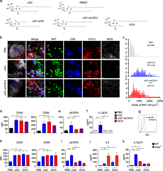
The spatiotemporal distribution of cytokines orchestrates immune responses in vivo, yet the underlying mechanisms remain to be explored. We showed here that the spatial distribution of interleukin-4 (IL4) in invariant natural killer T (iNKT) cells regulated crosstalk between iNKT cells and dendritic cells (DCs) and controlled iNKT cell-mediated T-helper type 1 (Th1) responses. The persistent polarization of IL4 induced by strong lipid antigens, that is, α-galactosylceramide (αGC), caused IL4 accumulation at the immunological synapse (IS), which promoted the activation of the IL4R-STAT6 (signal transducer and activator of transcription 6) pathway and production of IL12 in DCs, which enhanced interferon-γ (IFNγ) production in iNKT cells. Conversely, the nonpolarized secretion of IL4 induced by Th2 lipid antigens with a short or unsaturated chain was incapable of enhancing this iNKT cell-DC crosstalk and thus shifted the immune response to a Th2-type response. The nonpolarized secretion of IL4 in response to Th2 lipid antigens was caused by the degradation of Cdc42 in iNKT cells. Moreover, reduced Cdc42 expression was observed in tumor-infiltrating iNKT cells, which impaired IL4 polarization and disturbed iNKT cell-DC crosstalk in tumors.
中文翻译:

IL4的空间分布控制着肿瘤中iNKT细胞与DC的串扰。
细胞因子的时空分布可在体内协调免疫反应,但其潜在机制仍有待探索。我们在这里显示,不变的自然杀伤T(iNKT)细胞中白介素4(IL4)的空间分布调节了iNKT细胞与树突状细胞(DC)之间的串扰,并控制了iNKT细胞介导的1型T辅助反应(Th1)。强脂抗原即α-半乳糖基神经酰胺(αGC)诱导的IL4持续极化导致IL4在免疫突触(IS)处积聚,从而促进IL4R-STAT6的激活(信号转导子和转录激活子6)。 DCs中IL12的信号通路和产生,这增强了iNKT细胞中干扰素-γ(IFNγ)的产生。反过来,Th2脂抗原具有短链或不饱和链诱导的IL4的非极化分泌无法增强这种iNKT细胞-DC串扰,因此将免疫应答转变为Th2型应答。IL4对Th2脂质抗原的非极化分泌是由iNKT细胞中Cdc42的降解引起的。此外,在肿瘤浸润的iNKT细胞中观察到了Cdc42表达降低,这削弱了IL4极化并干扰了肿瘤中的iNKT细胞-DC串扰。
更新日期:2019-06-03
中文翻译:

IL4的空间分布控制着肿瘤中iNKT细胞与DC的串扰。
细胞因子的时空分布可在体内协调免疫反应,但其潜在机制仍有待探索。我们在这里显示,不变的自然杀伤T(iNKT)细胞中白介素4(IL4)的空间分布调节了iNKT细胞与树突状细胞(DC)之间的串扰,并控制了iNKT细胞介导的1型T辅助反应(Th1)。强脂抗原即α-半乳糖基神经酰胺(αGC)诱导的IL4持续极化导致IL4在免疫突触(IS)处积聚,从而促进IL4R-STAT6的激活(信号转导子和转录激活子6)。 DCs中IL12的信号通路和产生,这增强了iNKT细胞中干扰素-γ(IFNγ)的产生。反过来,Th2脂抗原具有短链或不饱和链诱导的IL4的非极化分泌无法增强这种iNKT细胞-DC串扰,因此将免疫应答转变为Th2型应答。IL4对Th2脂质抗原的非极化分泌是由iNKT细胞中Cdc42的降解引起的。此外,在肿瘤浸润的iNKT细胞中观察到了Cdc42表达降低,这削弱了IL4极化并干扰了肿瘤中的iNKT细胞-DC串扰。


 京公网安备 11010802027423号