Bone Research ( IF 14.3 ) Pub Date : 2019-05-22 , DOI: 10.1038/s41413-019-0053-z Furqan A Shah 1 , Krisztina Ruscsák 1 , Anders Palmquist 1

|
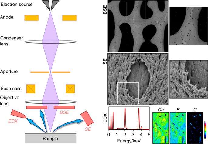
Bone is an architecturally complex system that constantly undergoes structural and functional optimisation through renewal and repair. The scanning electron microscope (SEM) is among the most frequently used instruments for examining bone. It offers the key advantage of very high spatial resolution coupled with a large depth of field and wide field of view. Interactions between incident electrons and atoms on the sample surface generate backscattered electrons, secondary electrons, and various other signals including X-rays that relay compositional and topographical information. Through selective removal or preservation of specific tissue components (organic, inorganic, cellular, vascular), their individual contribution(s) to the overall functional competence can be elucidated. With few restrictions on sample geometry and a variety of applicable sample-processing routes, a given sample may be conveniently adapted for multiple analytical methods. While a conventional SEM operates at high vacuum conditions that demand clean, dry, and electrically conductive samples, non-conductive materials (e.g., bone) can be imaged without significant modification from the natural state using an environmental scanning electron microscope. This review highlights important insights gained into bone microstructure and pathophysiology, bone response to implanted biomaterials, elemental analysis, SEM in paleoarchaeology, 3D imaging using focused ion beam techniques, correlative microscopy and in situ experiments. The capacity to image seamlessly across multiple length scales within the meso-micro-nano-continuum, the SEM lends itself to many unique and diverse applications, which attest to the versatility and user-friendly nature of this instrument for studying bone. Significant technological developments are anticipated for analysing bone using the SEM.
中文翻译:

骨扫描电子显微镜 50 年——对健康、疾病和埋藏学中骨材料特性的重要发现和见解的全面概述
骨骼是一个结构复杂的系统,通过更新和修复不断进行结构和功能优化。扫描电子显微镜 (SEM) 是最常用的骨骼检查仪器之一。它提供了非常高的空间分辨率以及大景深和宽视场的关键优势。入射电子和样品表面原子之间的相互作用产生背散射电子、二次电子和各种其他信号,包括传递成分和形貌信息的 X 射线。通过选择性去除或保留特定组织成分(有机、无机、细胞、血管),可以阐明它们对整体功能能力的个体贡献。由于对样品几何形状和各种适用的样品处理路线的限制很少,给定的样品可以方便地适应多种分析方法。虽然传统的 SEM 在高真空条件下运行,需要清洁、干燥和导电的样品,但使用环境扫描电子显微镜可以对非导电材料(例如骨骼)进行成像,而无需对自然状态进行显着修改。这篇综述重点介绍了对骨微观结构和病理生理学、骨对植入生物材料的反应、元素分析、古考古学中的 SEM、使用聚焦离子束技术的 3D 成像、相关显微镜和原位实验的重要见解。SEM 能够在介观-微-纳米连续体内的多个长度尺度上无缝成像,使其适合许多独特且多样化的应用,这证明了该仪器用于研究骨骼的多功能性和用户友好性。使用 SEM 分析骨骼预计将出现重大技术发展。


 京公网安备 11010802027423号