当前位置:
X-MOL 学术
›
Microchim. Acta
›
论文详情
Our official English website, www.x-mol.net, welcomes your
feedback! (Note: you will need to create a separate account there.)
Distinguishing cancer cell lines at a single living cell level via detection of sialic acid by dual-channel plasmonic imaging and by using a SERS-microfluidic droplet platform
Microchimica Acta ( IF 5.3 ) Pub Date : 2019-05-21 , DOI: 10.1007/s00604-019-3480-z Lili Cong , Lijia Liang , Fanghao Cao , Dan Sun , Jing Yue , Weiqing Xu , Chongyang Liang , Shuping Xu
Microchimica Acta ( IF 5.3 ) Pub Date : 2019-05-21 , DOI: 10.1007/s00604-019-3480-z Lili Cong , Lijia Liang , Fanghao Cao , Dan Sun , Jing Yue , Weiqing Xu , Chongyang Liang , Shuping Xu
|
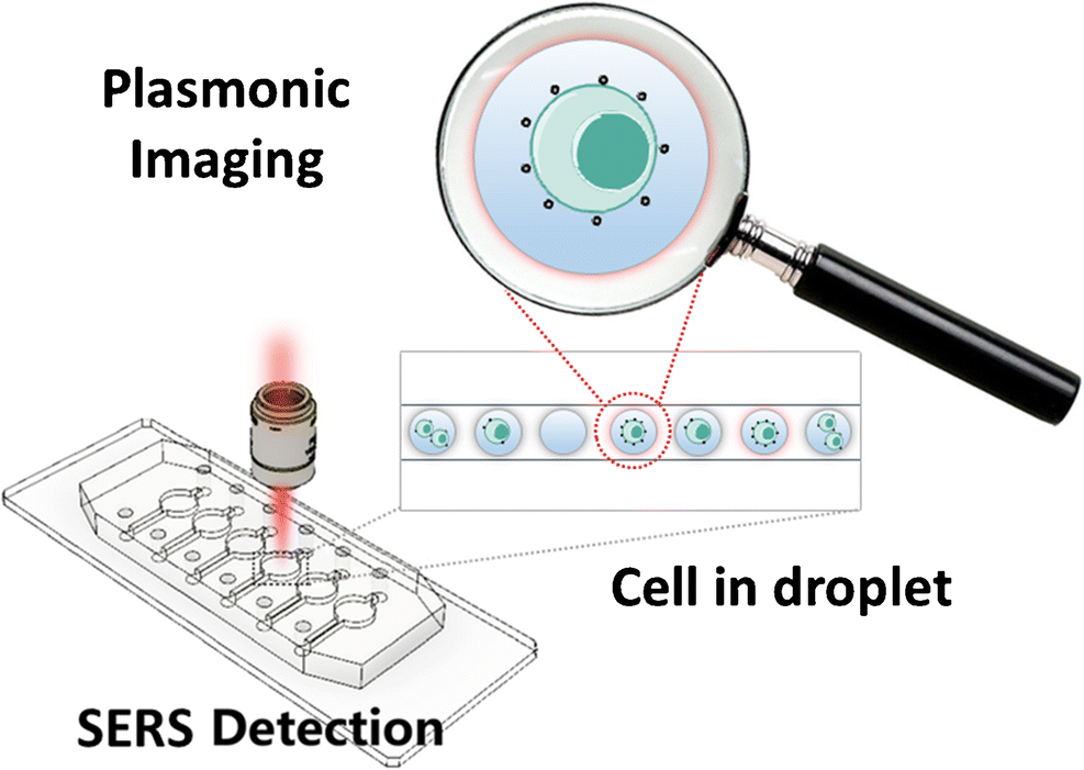
AbstractA high-throughput, dual-channel single cell analytical method is described for the detection of sialic acid (SA) on single cell based on the use of microfluidic droplets integrated with plasmonic imaging and surface-enhanced Raman spectroscopy (SERS) with the assistance of a multifunctional metal nanoparticle-based probe. The multifunctional plasmonic nanoprobe was prepared by modifying silver nanoparticles (AgNPs) with 4-mercaptophenylboronic acid (MPBA) that both warrants SA recognition and acts as a Raman reporter. This nanoprobe is a high-contrast indicator under bright field imaging due to the strong energy loss feature of AgNPs, and also owns possesses a strong SERS enhancement capability toward MPBA. Cells incubated with the plasmonic nanoprobes were isolated in water-in-oil droplets and then were re-dispersed in a chamber array chip. High-precision profiles of SA on a single cell in one droplet were obtained by the bright field imaging and image processing. The SA expression levels on different cell lines (MCF-7, HepG2, SGC and BNL.CL2) traced by SERS spectroscopy were compared. The statistical data among different cell lines confirm that the SA expression levels on cancer cells are much higher than that on normal cells. Single cell analysis further revealed that the cell-to-cell variations are more obvious in cancer cell lines. This study provides a valuable tool for understanding glycan-related biochemical processes. Graphical abstractA high-throughput, dual-channel microfluidic droplet platform succeeded in distinguishing different cancer cell lines at single living cell level integrated with plasmonic imaging and surface-enhanced Raman spectroscopy with assistance of a multifunctional metal nanoparticle-based probe.
中文翻译:

通过双通道等离子体成像和使用 SERS 微流体液滴平台检测唾液酸在单个活细胞水平上区分癌细胞系
摘要描述了一种高通量、双通道单细胞分析方法,基于使用微流体液滴与等离子体成像和表面增强拉曼光谱 (SERS) 相结合,在单细胞上检测唾液酸 (SA)。一种多功能金属纳米颗粒探针。多功能等离子体纳米探针是通过用 4-巯基苯基硼酸 (MPBA) 修饰银纳米粒子 (AgNPs) 制备的,该纳米粒子既保证 SA 识别,又充当拉曼报告分子。由于AgNPs的强能量损失特性,该纳米探针是明场成像下的高对比度指示剂,并且对MPBA具有很强的SERS增强能力。用等离子体纳米探针孵育的细胞在油包水液滴中分离,然后重新分散在室阵列芯片中。通过明场成像和图像处理获得了一滴中单个细胞上SA的高精度轮廓。比较了通过 SERS 光谱追踪的不同细胞系(MCF-7、HepG2、SGC 和 BNL.CL2)上的 SA 表达水平。不同细胞系之间的统计数据证实,癌细胞上的SA表达水平远高于正常细胞上的表达水平。单细胞分析进一步显示,癌细胞系中细胞间变异更为明显。这项研究为理解与聚糖相关的生化过程提供了宝贵的工具。图文摘要A高通量,
更新日期:2019-05-21
中文翻译:
通过双通道等离子体成像和使用 SERS 微流体液滴平台检测唾液酸在单个活细胞水平上区分癌细胞系
摘要描述了一种高通量、双通道单细胞分析方法,基于使用微流体液滴与等离子体成像和表面增强拉曼光谱 (SERS) 相结合,在单细胞上检测唾液酸 (SA)。一种多功能金属纳米颗粒探针。多功能等离子体纳米探针是通过用 4-巯基苯基硼酸 (MPBA) 修饰银纳米粒子 (AgNPs) 制备的,该纳米粒子既保证 SA 识别,又充当拉曼报告分子。由于AgNPs的强能量损失特性,该纳米探针是明场成像下的高对比度指示剂,并且对MPBA具有很强的SERS增强能力。用等离子体纳米探针孵育的细胞在油包水液滴中分离,然后重新分散在室阵列芯片中。通过明场成像和图像处理获得了一滴中单个细胞上SA的高精度轮廓。比较了通过 SERS 光谱追踪的不同细胞系(MCF-7、HepG2、SGC 和 BNL.CL2)上的 SA 表达水平。不同细胞系之间的统计数据证实,癌细胞上的SA表达水平远高于正常细胞上的表达水平。单细胞分析进一步显示,癌细胞系中细胞间变异更为明显。这项研究为理解与聚糖相关的生化过程提供了宝贵的工具。图文摘要A高通量,


 京公网安备 11010802027423号