当前位置:
X-MOL 学术
›
J. Hepatol.
›
论文详情
Our official English website, www.x-mol.net, welcomes your
feedback! (Note: you will need to create a separate account there.)
Arterial Subtraction Images of Gadoxetate-Enhanced MRI Improve Diagnosis of Early-Stage Hepatocellular Carcinoma
Journal of Hepatology ( IF 26.8 ) Pub Date : 2019-09-01 , DOI: 10.1016/j.jhep.2019.05.005 Dong Hwan Kim 1 , Sang Hyun Choi 1 , Jae Ho Byun 1 , Ji Hun Kang 1 , Young-Suk Lim 2 , So Jung Lee 1 , So Yeon Kim 1 , Hyung Jin Won 1 , Yong Moon Shin 1 , Pyo-Nyun Kim 1
Journal of Hepatology ( IF 26.8 ) Pub Date : 2019-09-01 , DOI: 10.1016/j.jhep.2019.05.005 Dong Hwan Kim 1 , Sang Hyun Choi 1 , Jae Ho Byun 1 , Ji Hun Kang 1 , Young-Suk Lim 2 , So Jung Lee 1 , So Yeon Kim 1 , Hyung Jin Won 1 , Yong Moon Shin 1 , Pyo-Nyun Kim 1
Affiliation
|
BACKGROUND & AIMS
Although gadoxetate disodium-enhanced magnetic resonance imaging (MRI) shows higher sensitivity for diagnosing hepatocellular carcinoma (HCC), its arterial-phase images may be unsatisfactory because of weak arterial enhancement. We investigated the clinical effectiveness of arterial subtraction images from gadoxetate disodium-enhanced MRI for diagnosing early-stage HCC using the Liver Imaging Reporting and Data System (LI-RADS) v2018. METHODS
A total of 372 hepatic nodules (273 HCCs, 18 other malignancies, and 81 benign nodules) of 3.0 cm or smaller from 258 patients at risk of HCC who underwent gadoxetate disodium-enhanced MRI in 2016 were retrospectively analyzed. Final diagnosis was assessed histopathologically or clinically (marginal recurrence after treatment or change in lesion size on follow-up imaging). The detection rate for arterial hyperenhancement was compared between ordinary arterial-phase and arterial subtraction images, and the benefit of arterial subtraction images in diagnosing HCC using LI-RADS was assessed. RESULTS
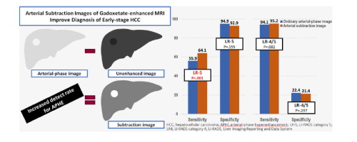
Arterial subtraction images had a significantly higher detection rate for arterial hyperenhancement than ordinary arterial-phase images, both for all hepatic nodules (72.3% vs. 62.4%, p<.001) and HCCs (91.9% vs. 80.6%, p<.001). Compared with ordinary arterial-phase images, arterial subtraction images significantly increased the sensitivity of LI-RADS category 5 for diagnosis of HCC (64.1% [173/270] vs. 55.9% [151/270], p<.001), without significantly decreasing specificity (92.9% [91/98] vs. 94.9% [93/98], p=.155). For histopathologically confirmed lesions, arterial subtraction images significantly increased sensitivity to 68.8% (128/186) from the 61.3% (114/186) of ordinary arterial-phase images (p<.001), with a minimal decrease in specificity to 84.8% (39/46) from 89.1% (41/46) (p=.151). CONCLUSIONS
Arterial subtraction images of gadoxetate disodium-enhanced MRI can significantly improve the sensitivity for diagnosing early-stage HCC, without a significant decrease in specificity, using LI-RADS. LAY SUMMARY
When applied to the LI-RADS on gadoxetate disodium-enhanced MRI, the arterial subtraction images are clinically useful to improve diagnosis of early-stage HCC.
中文翻译:

Gadoxetate增强MRI的动脉减影图像改善早期肝细胞癌的诊断
背景与目的虽然钆塞酸二钠增强磁共振成像(MRI)对诊断肝细胞癌(HCC)具有较高的敏感性,但由于动脉增强较弱,其动脉期图像可能并不令人满意。我们使用肝脏影像报告和数据系统 (LI-RADS) v2018 研究了钆塞酸二钠增强 MRI 的动脉减影图像诊断早期 HCC 的临床有效性。方法 对2016年接受钆塞酸二钠增强MRI检查的258例有HCC风险的患者共372个肝结节(273个HCC、18个其他恶性肿瘤和81个良性结节)进行了回顾性分析。通过组织病理学或临床评估最终诊断(治疗后边缘复发或随访影像学病变大小变化)。比较了普通动脉期和动脉减影影像对动脉超增强的检出率,并评估了动脉减影影像在使用 LI-RADS 诊断 HCC 中的益处。结果对于所有肝结节(72.3% 对 62.4%,p<.001)和 HCC(91.9% 对 80.6%,p< .001)。与普通动脉期图像相比,动脉减影图像显着提高了 LI-RADS 5 类诊断 HCC 的敏感性(64.1% [173/270] vs. 55.9% [151/270],p<.001),而没有特异性显着降低(92.9% [91/98] vs. 94.9% [93/98],p=.155)。对于组织病理学证实的病变,动脉减影图像的敏感性从普通动脉期图像的 61.3% (114/186) 显着提高至 68.8% (128/186) (p<.001),特异性最低降至 84.8% (39/46)从 89.1% (41/46) (p=.151)。结论钆塞酸二钠增强 MRI 的动脉减影图像可以显着提高使用 LI-RADS 诊断早期 HCC 的敏感性,而没有显着降低特异性。常规总结 当应用于钆塞酸二钠增强 MRI 上的 LI-RADS 时,动脉减影图像在临床上可用于改善早期 HCC 的诊断。结论钆塞酸二钠增强 MRI 的动脉减影图像可以显着提高使用 LI-RADS 诊断早期 HCC 的敏感性,而没有显着降低特异性。常规总结 当应用于钆塞酸二钠增强 MRI 上的 LI-RADS 时,动脉减影图像在临床上可用于改善早期 HCC 的诊断。结论钆塞酸二钠增强 MRI 的动脉减影图像可以显着提高使用 LI-RADS 诊断早期 HCC 的敏感性,而没有显着降低特异性。常规总结 当应用于钆塞酸二钠增强 MRI 上的 LI-RADS 时,动脉减影图像在临床上可用于改善早期 HCC 的诊断。
更新日期:2019-09-01
中文翻译:
Gadoxetate增强MRI的动脉减影图像改善早期肝细胞癌的诊断
背景与目的虽然钆塞酸二钠增强磁共振成像(MRI)对诊断肝细胞癌(HCC)具有较高的敏感性,但由于动脉增强较弱,其动脉期图像可能并不令人满意。我们使用肝脏影像报告和数据系统 (LI-RADS) v2018 研究了钆塞酸二钠增强 MRI 的动脉减影图像诊断早期 HCC 的临床有效性。方法 对2016年接受钆塞酸二钠增强MRI检查的258例有HCC风险的患者共372个肝结节(273个HCC、18个其他恶性肿瘤和81个良性结节)进行了回顾性分析。通过组织病理学或临床评估最终诊断(治疗后边缘复发或随访影像学病变大小变化)。比较了普通动脉期和动脉减影影像对动脉超增强的检出率,并评估了动脉减影影像在使用 LI-RADS 诊断 HCC 中的益处。结果对于所有肝结节(72.3% 对 62.4%,p<.001)和 HCC(91.9% 对 80.6%,p< .001)。与普通动脉期图像相比,动脉减影图像显着提高了 LI-RADS 5 类诊断 HCC 的敏感性(64.1% [173/270] vs. 55.9% [151/270],p<.001),而没有特异性显着降低(92.9% [91/98] vs. 94.9% [93/98],p=.155)。对于组织病理学证实的病变,动脉减影图像的敏感性从普通动脉期图像的 61.3% (114/186) 显着提高至 68.8% (128/186) (p<.001),特异性最低降至 84.8% (39/46)从 89.1% (41/46) (p=.151)。结论钆塞酸二钠增强 MRI 的动脉减影图像可以显着提高使用 LI-RADS 诊断早期 HCC 的敏感性,而没有显着降低特异性。常规总结 当应用于钆塞酸二钠增强 MRI 上的 LI-RADS 时,动脉减影图像在临床上可用于改善早期 HCC 的诊断。结论钆塞酸二钠增强 MRI 的动脉减影图像可以显着提高使用 LI-RADS 诊断早期 HCC 的敏感性,而没有显着降低特异性。常规总结 当应用于钆塞酸二钠增强 MRI 上的 LI-RADS 时,动脉减影图像在临床上可用于改善早期 HCC 的诊断。结论钆塞酸二钠增强 MRI 的动脉减影图像可以显着提高使用 LI-RADS 诊断早期 HCC 的敏感性,而没有显着降低特异性。常规总结 当应用于钆塞酸二钠增强 MRI 上的 LI-RADS 时,动脉减影图像在临床上可用于改善早期 HCC 的诊断。


 京公网安备 11010802027423号