当前位置:
X-MOL 学术
›
Nano Lett.
›
论文详情
Our official English website, www.x-mol.net, welcomes your
feedback! (Note: you will need to create a separate account there.)
Spatially Resolving Lithiation in Silicon-Graphite Composite Electrodes via in Situ High-Energy X-ray Diffraction Computed Tomography.
Nano Letters ( IF 9.6 ) Pub Date : 2019-05-24 , DOI: 10.1021/acs.nanolett.9b00955 Donal P Finegan 1 , Antonis Vamvakeros 2, 3 , Lei Cao 1 , Chun Tan 4, 5 , Thomas M M Heenan 4, 5 , Sohrab R Daemi 4 , Simon D M Jacques 3 , Andrew M Beale 3, 6, 7 , Marco Di Michiel 2 , Kandler Smith 1 , Dan J L Brett 4, 5 , Paul R Shearing 4, 5 , Chunmei Ban 1
Nano Letters ( IF 9.6 ) Pub Date : 2019-05-24 , DOI: 10.1021/acs.nanolett.9b00955 Donal P Finegan 1 , Antonis Vamvakeros 2, 3 , Lei Cao 1 , Chun Tan 4, 5 , Thomas M M Heenan 4, 5 , Sohrab R Daemi 4 , Simon D M Jacques 3 , Andrew M Beale 3, 6, 7 , Marco Di Michiel 2 , Kandler Smith 1 , Dan J L Brett 4, 5 , Paul R Shearing 4, 5 , Chunmei Ban 1
Affiliation

|
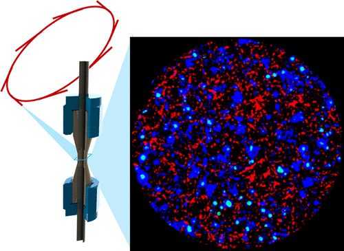
Optimizing the chemical and morphological parameters of lithium-ion (Li-ion) electrodes is extremely challenging, due in part to the absence of techniques to construct spatial and temporal descriptions of chemical and morphological heterogeneities. We present the first demonstration of combined high-speed X-ray diffraction (XRD) and XRD computed tomography (XRD-CT) to probe, in 3D, crystallographic heterogeneities within Li-ion electrodes with a spatial resolution of 1 μm. The local charge-transfer mechanism within and between individual particles was investigated in a silicon(Si)-graphite composite electrode. High-speed XRD revealed charge balancing kinetics between the graphite and Si during the minutes following the transition from operation to open circuit. Subparticle lithiation heterogeneities in both Si and graphite were observed using XRD-CT, where the core and shell structures were segmented, and their respective diffraction patterns were characterized.
中文翻译:

通过原位高能X射线衍射计算机断层扫描技术在硅石墨复合电极中空间分辨锂化。
优化锂离子(Li-ion)电极的化学和形态参数是极富挑战性的,部分原因是缺乏用于构造化学和形态异质性的时空描述的技术。我们目前首次展示了结合高速X射线衍射(XRD)和XRD计算机断层扫描(XRD-CT)的3D模式,以3D探测空间分辨率为1μm的锂离子电极内的结晶异质性。在硅(Si)-石墨复合电极中研究了单个颗粒内部和之间的局部电荷转移机理。高速XRD显示了从操作到开路过渡后的几分钟内,石墨和Si之间的电荷平衡动力学。
更新日期:2019-05-13
中文翻译:

通过原位高能X射线衍射计算机断层扫描技术在硅石墨复合电极中空间分辨锂化。
优化锂离子(Li-ion)电极的化学和形态参数是极富挑战性的,部分原因是缺乏用于构造化学和形态异质性的时空描述的技术。我们目前首次展示了结合高速X射线衍射(XRD)和XRD计算机断层扫描(XRD-CT)的3D模式,以3D探测空间分辨率为1μm的锂离子电极内的结晶异质性。在硅(Si)-石墨复合电极中研究了单个颗粒内部和之间的局部电荷转移机理。高速XRD显示了从操作到开路过渡后的几分钟内,石墨和Si之间的电荷平衡动力学。


 京公网安备 11010802027423号