Nature Protocols ( IF 13.1 ) Pub Date : 2019-05-03 , DOI: 10.1038/s41596-019-0160-8 Johanna F. Dekkers , Maria Alieva , Lianne M. Wellens , Hendrikus C. R. Ariese , Paul R. Jamieson , Annelotte M. Vonk , Gimano D. Amatngalim , Huili Hu , Koen C. Oost , Hugo J. G. Snippert , Jeffrey M. Beekman , Ellen J. Wehrens , Jane E. Visvader , Hans Clevers , Anne C. Rios
|
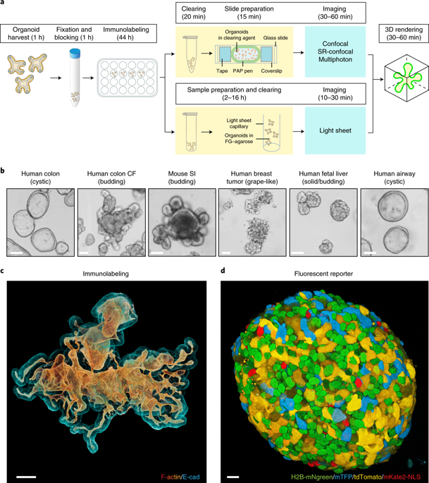
In vitro 3D organoid systems have revolutionized the modeling of organ development and diseases in a dish. Fluorescence microscopy has contributed to the characterization of the cellular composition of organoids and demonstrated organoids’ phenotypic resemblance to their original tissues. Here, we provide a detailed protocol for performing high-resolution 3D imaging of entire organoids harboring fluorescence reporters and upon immunolabeling. This method is applicable to a wide range of organoids of differing origins and of various sizes and shapes. We have successfully used it on human airway, colon, kidney, liver and breast tumor organoids, as well as on mouse mammary gland organoids. It includes a simple clearing method utilizing a homemade fructose–glycerol clearing agent that captures 3D organoids in full and enables marker quantification on a cell-by-cell basis. Sample preparation has been optimized for 3D imaging by confocal, super-resolution confocal, multiphoton and light-sheet microscopy. From organoid harvest to image analysis, the protocol takes 3 d.
中文翻译:
固定和清除的类动物体的高分辨率3D成像
体外3D类器官系统彻底改变了器皿中器官发育和疾病的建模。荧光显微镜有助于类器官的细胞组成的表征,并证明类器官的表型与其原始组织相似。在这里,我们提供了一个详细的协议,可对带有荧光报告分子的整个类器官进行高分辨率3D成像,并进行免疫标记。该方法适用于各种起源不同,大小和形状各异的类器官。我们已经成功地将其用于人的气道,结肠,肾脏,肝脏和乳腺肿瘤的类器官,以及小鼠的乳腺类器官。它包括一种简单的清除方法,该方法利用自制的果糖-甘油清除剂,可以完全捕获3D类器官,并可以逐个细胞地进行标记定量。通过共聚焦,超分辨率共聚焦,多光子和光片显微镜,样品制备已针对3D成像进行了优化。从类器官收获到图像分析,该协议需要3 d。


 京公网安备 11010802027423号