当前位置:
X-MOL 学术
›
Anal. Chem.
›
论文详情
Our official English website, www.x-mol.net, welcomes your
feedback! (Note: you will need to create a separate account there.)
α-Synuclein-Confocal Nanoscanning (ASYN-CONA), a Bead-Based Assay for Detecting Early-Stage α-Synuclein Aggregation.
Analytical Chemistry ( IF 6.7 ) Pub Date : 2019-04-18 , DOI: 10.1021/acs.analchem.8b03842 Irene Pérez-Pi 1 , David A Evans 1 , Mathew H Horrocks 2, 3 , Nhan T Pham 1 , Karamjit S Dolt 4 , Joanna Koszela 1 , Tilo Kunath 4 , Manfred Auer 1
Analytical Chemistry ( IF 6.7 ) Pub Date : 2019-04-18 , DOI: 10.1021/acs.analchem.8b03842 Irene Pérez-Pi 1 , David A Evans 1 , Mathew H Horrocks 2, 3 , Nhan T Pham 1 , Karamjit S Dolt 4 , Joanna Koszela 1 , Tilo Kunath 4 , Manfred Auer 1
Affiliation
|
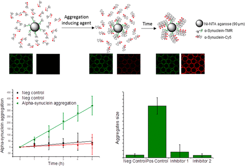
α-Synuclein fibrils are considered a hallmark of Parkinson's disease and other synucleinopathies. However, small oligomers that formed during the early stages of α-synuclein aggregation are thought to be the main toxic species causing disease. The formation of α-synuclein oligomers has proven difficult to follow, because of the heterogeneity and transient nature of the species formed. Here, a novel bead-based aggregation assay for monitoring the earliest stages of α-synuclein oligomerization, α-Synuclein-Confocal Nanoscanning (ASYN-CONA), is presented. The α-synuclein A91C single cysteine mutant is modified with a trifunctional chemical tag, which allows simultaneous fluorescent labeling with a green dye (tetramethylrhodamine, TMR) and attachment to microbeads. Beads with bound TMR-labeled α-synuclein are then incubated with a red dye (Cy5)-labeled variant of α-synuclein A91C, and EtOH (20%) to induce aggregation. Aggregation is detected by confocal scanning imaging, below the equatorial plane of the beads, which is known as the CONA technique. On-bead TMR-labeled α-synuclein and aggregated Cy5-labeled α-synuclein from the solution are quantitatively monitored in parallel by detection of fluorescent halos or "rings". α-Synuclein on-bead oligomerization results in a linear increase of red bead ring fluorescence intensity over a period of 5 h. Total internal reflection fluorescence microscopy was performed on oligomers cleaved from the beads, and it revealed that (i) oligomers are sufficiently stable in solution to investigate their composition, consisting of 6 ± 1 monomer units, and (ii) oligomers containing a mean of 15 monomers bind Thioflavin-T. Various known inhibitors of α-synuclein aggregation were used to validate the ASYN-CONA assay for drug screening. Baicalein, curcumin, and rifampicin showed concentration-dependent inhibition of the α-synuclein aggregation and the IC50 (the concentration of the compound at which the maxiumum intensity was reduced by one-half) were calculated.
中文翻译:

α-突触核蛋白共聚焦纳米扫描 (ASYN-CONA),一种用于检测早期 α-突触核蛋白聚集的基于微珠的测定。
α-突触核蛋白原纤维被认为是帕金森病和其他突触核蛋白病的标志。然而,在 α-突触核蛋白聚集的早期阶段形成的小寡聚体被认为是导致疾病的主要有毒物质。由于所形成物种的异质性和短暂性,α-突触核蛋白寡聚体的形成已被证明难以遵循。在这里,提出了一种新的基于珠子的聚集测定法,用于监测 α-突触核蛋白寡聚化的最早阶段,α-突触核蛋白-共聚焦纳米扫描 (ASYN-CONA)。α-突触核蛋白 A91C 单半胱氨酸突变体经过三功能化学标签修饰,可同时使用绿色染料(四甲基罗丹明,TMR)进行荧光标记并附着在微珠上。然后将具有结合 TMR 标记的 α-突触核蛋白的珠与红色染料 (Cy5) 标记的 α-突触核蛋白 A91C 变体和 EtOH (20%) 一起孵育以诱导聚集。通过共焦扫描成像在珠子的赤道平面下方检测聚集,这被称为 CONA 技术。通过检测荧光晕圈或“环”,并行定量监测溶液中的珠上 TMR 标记的 α-突触核蛋白和聚集的 Cy5 标记的 α-突触核蛋白。α-突触核蛋白珠上寡聚导致红珠环荧光强度在 5 小时内线性增加。对从珠子上切割下来的低聚物进行全内反射荧光显微镜检查,结果表明(i)低聚物在溶液中足够稳定,可以研究它们的组成,由 6 ± 1 个单体单元组成,(ii) 平均含有 15 个单体的低聚物结合硫黄素-T。各种已知的 α-突触核蛋白聚集抑制剂被用于验证用于药物筛选的 ASYN-CONA 测定。黄芩素、姜黄素和利福平对α-突触核蛋白聚集具有浓度依赖性抑制作用,计算出IC50(最大强度降低二分之一时的化合物浓度)。
更新日期:2019-04-09
中文翻译:
α-突触核蛋白共聚焦纳米扫描 (ASYN-CONA),一种用于检测早期 α-突触核蛋白聚集的基于微珠的测定。
α-突触核蛋白原纤维被认为是帕金森病和其他突触核蛋白病的标志。然而,在 α-突触核蛋白聚集的早期阶段形成的小寡聚体被认为是导致疾病的主要有毒物质。由于所形成物种的异质性和短暂性,α-突触核蛋白寡聚体的形成已被证明难以遵循。在这里,提出了一种新的基于珠子的聚集测定法,用于监测 α-突触核蛋白寡聚化的最早阶段,α-突触核蛋白-共聚焦纳米扫描 (ASYN-CONA)。α-突触核蛋白 A91C 单半胱氨酸突变体经过三功能化学标签修饰,可同时使用绿色染料(四甲基罗丹明,TMR)进行荧光标记并附着在微珠上。然后将具有结合 TMR 标记的 α-突触核蛋白的珠与红色染料 (Cy5) 标记的 α-突触核蛋白 A91C 变体和 EtOH (20%) 一起孵育以诱导聚集。通过共焦扫描成像在珠子的赤道平面下方检测聚集,这被称为 CONA 技术。通过检测荧光晕圈或“环”,并行定量监测溶液中的珠上 TMR 标记的 α-突触核蛋白和聚集的 Cy5 标记的 α-突触核蛋白。α-突触核蛋白珠上寡聚导致红珠环荧光强度在 5 小时内线性增加。对从珠子上切割下来的低聚物进行全内反射荧光显微镜检查,结果表明(i)低聚物在溶液中足够稳定,可以研究它们的组成,由 6 ± 1 个单体单元组成,(ii) 平均含有 15 个单体的低聚物结合硫黄素-T。各种已知的 α-突触核蛋白聚集抑制剂被用于验证用于药物筛选的 ASYN-CONA 测定。黄芩素、姜黄素和利福平对α-突触核蛋白聚集具有浓度依赖性抑制作用,计算出IC50(最大强度降低二分之一时的化合物浓度)。


 京公网安备 11010802027423号