Nature Reviews Gastroenterology & Hepatology ( IF 45.9 ) Pub Date : 2019-04-01 , DOI: 10.1038/s41575-019-0134-x
Shani Ben-Moshe 1 , Shalev Itzkovitz 1

|
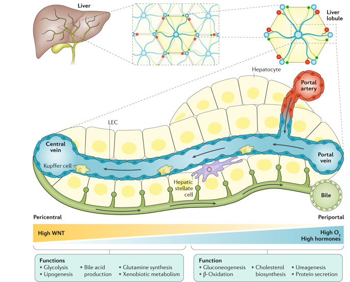
Hepatocytes operate in highly structured repeating anatomical units termed liver lobules. Blood flow along the lobule radial axis creates gradients of oxygen, nutrients and hormones, which, together with morphogenetic fields, give rise to a highly variable microenvironment. In line with this spatial variability, key liver functions are expressed non-uniformly across the lobules, a phenomenon termed zonation. Technologies based on single-cell transcriptomics have constructed a global spatial map of hepatocyte gene expression in mice revealing that ~50% of hepatocyte genes are expressed in a zonated manner. This broad spatial heterogeneity suggests that hepatocytes in different lobule zones might have not only different gene expression profiles but also distinct epigenetic features, regenerative capacities, susceptibilities to damage and other functional aspects. Here, we present genomic approaches for studying liver zonation, describe the principles of liver zonation and discuss the intrinsic and extrinsic factors that dictate zonation patterns. We also explore the challenges and solutions for obtaining zonation maps of liver non-parenchymal cells. These approaches facilitate global characterization of liver function with high spatial resolution along physiological and pathological timescales.
中文翻译:

哺乳动物肝脏的空间异质性
肝细胞在称为肝小叶的高度结构化的重复解剖单元中运行。沿着小叶径向轴的血流产生了氧气、营养物质和激素的梯度,这些梯度与形态发生场一起产生了高度可变的微环境。与这种空间变异性一致,关键肝功能在小叶中的表达不均匀,这种现象称为分区。基于单细胞转录组学的技术已经构建了小鼠肝细胞基因表达的全球空间图,揭示了约 50% 的肝细胞基因以分区方式表达。这种广泛的空间异质性表明,不同小叶区域的肝细胞可能不仅具有不同的基因表达谱,而且还具有不同的表观遗传特征、再生能力、对损坏和其他功能方面的敏感性。在这里,我们提出了研究肝脏分区的基因组方法,描述了肝脏分区的原理,并讨论了决定分区模式的内在和外在因素。我们还探讨了获得肝脏非实质细胞分区图的挑战和解决方案。这些方法有助于沿生理和病理时间尺度以高空间分辨率对肝功能进行全局表征。


 京公网安备 11010802027423号