Scientific Reports ( IF 3.8 ) Pub Date : 2019-03-14 , DOI: 10.1038/s41598-019-38813-2 Mahmoud Abdolhoseini , Murielle G. Kluge , Frederick R. Walker , Sarah J. Johnson

|
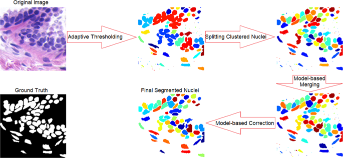
Automated cell nucleus segmentation is the key to gain further insight into cell features and functionality which support computer-aided pathology in early diagnosis of diseases such as breast cancer and brain tumour. Despite considerable advances in automated segmentation, it still remains a challenging task to split heavily clustered nuclei due to intensity variations caused by noise and uneven absorption of stains. To address this problem, we propose a novel method applicable to variety of histopathological images stained for different proteins, with high speed, accuracy and level of automation. Our algorithm is initiated by applying a new locally adaptive thresholding method on watershed regions. Followed by a new splitting technique based on multilevel thresholding and the watershed algorithm to separate clustered nuclei. Finalized by a model-based merging step to eliminate oversegmentation and a model-based correction step to improve segmentation results and eliminate small objects. We have applied our method to three image datasets: breast cancer stained for hematoxylin and eosin (H&E), Drosophila Kc167 cells stained for DNA to label nuclei, and mature neurons stained for NeuN. Evaluated results show our method outperforms the state-of-the-art methods in terms of accuracy, precision, F1-measure, and computational time.
中文翻译:

从组织病理学图像分割重聚集的细胞核
自动化的细胞核分割是进一步了解细胞特征和功能的关键,这些特征和功能支持计算机辅助病理学,以早期诊断诸如乳腺癌和脑瘤之类的疾病。尽管在自动分割方面取得了长足的进步,但由于噪声和污渍吸收不均匀引起的强度变化,分裂重聚集的核仍然是一项艰巨的任务。为了解决这个问题,我们提出了一种新颖的方法,该方法适用于对不同蛋白质染色的各种组织病理学图像,具有很高的速度,准确性和自动化程度。我们的算法是通过在分水岭地区应用一种新的局部自适应阈值方法来启动的。随后是基于多级阈值和分水岭算法的新分裂技术,以分离簇状核。通过基于模型的合并步骤(以消除过度分割)和基于模型的校正步骤(以改善分割结果并消除小物体)完成。我们已将我们的方法应用于三个图像数据集:对苏木精和曙红(H&E)染色的乳腺癌,对DNA标记核的果蝇Kc167细胞进行染色,对NeuN进行染色的成熟神经元。评估结果表明,我们的方法在准确性,精度,F1度量和计算时间方面均优于最新方法。


 京公网安备 11010802027423号