当前位置:
X-MOL 学术
›
Nat. Nanotechnol.
›
论文详情
Our official English website, www.x-mol.net, welcomes your
feedback! (Note: you will need to create a separate account there.)
Bright sub-20-nm cathodoluminescent nanoprobes for electron microscopy.
Nature Nanotechnology ( IF 38.1 ) Pub Date : 2019-03-04 , DOI: 10.1038/s41565-019-0395-0 Maxim B Prigozhin 1 , Peter C Maurer 1 , Alexandra M Courtis 2 , Nian Liu 3, 4 , Michael D Wisser 3 , Chris Siefe 3 , Bining Tian 5 , Emory Chan 5 , Guosheng Song 6, 7 , Stefan Fischer 3 , Shaul Aloni 5 , D Frank Ogletree 5 , Edward S Barnard 5 , Lydia-Marie Joubert 8, 9 , Jianghong Rao 6 , A Paul Alivisatos 2, 10, 11, 12 , Roger M Macfarlane 13 , Bruce E Cohen 5 , Yi Cui 3 , Jennifer A Dionne 3 , Steven Chu 1, 14
Nature Nanotechnology ( IF 38.1 ) Pub Date : 2019-03-04 , DOI: 10.1038/s41565-019-0395-0 Maxim B Prigozhin 1 , Peter C Maurer 1 , Alexandra M Courtis 2 , Nian Liu 3, 4 , Michael D Wisser 3 , Chris Siefe 3 , Bining Tian 5 , Emory Chan 5 , Guosheng Song 6, 7 , Stefan Fischer 3 , Shaul Aloni 5 , D Frank Ogletree 5 , Edward S Barnard 5 , Lydia-Marie Joubert 8, 9 , Jianghong Rao 6 , A Paul Alivisatos 2, 10, 11, 12 , Roger M Macfarlane 13 , Bruce E Cohen 5 , Yi Cui 3 , Jennifer A Dionne 3 , Steven Chu 1, 14
Affiliation

|
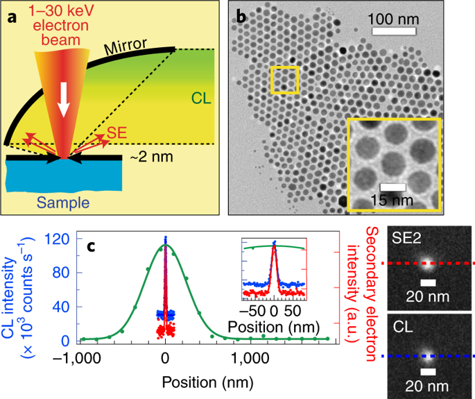
Electron microscopy has been instrumental in our understanding of complex biological systems. Although electron microscopy reveals cellular morphology with nanoscale resolution, it does not provide information on the location of different types of proteins. An electron-microscopy-based bioimaging technology capable of localizing individual proteins and resolving protein-protein interactions with respect to cellular ultrastructure would provide important insights into the molecular biology of a cell. Here, we synthesize small lanthanide-doped nanoparticles and measure the absolute photon emission rate of individual nanoparticles resulting from a given electron excitation flux (cathodoluminescence). Our results suggest that the optimization of nanoparticle composition, synthesis protocols and electron imaging conditions can lead to sub-20-nm nanolabels that would enable high signal-to-noise localization of individual biomolecules within a cellular context. In ensemble measurements, these labels exhibit narrow spectra of nine distinct colours, so the imaging of biomolecules in a multicolour electron microscopy modality may be possible.
中文翻译:

用于电子显微镜的20纳米以下亮阴极发光纳米探针。
电子显微镜对我们了解复杂的生物系统很有帮助。尽管电子显微镜揭示了具有纳米级分辨率的细胞形态,但它并未提供有关不同类型蛋白质位置的信息。基于电子显微镜的生物成像技术能够定位单个蛋白质并解决蛋白质-蛋白质在细胞超微结构方面的相互作用,这将为细胞分子生物学提供重要的见识。在这里,我们合成了镧系元素掺杂的纳米粒子,并测量了由给定的电子激发通量(阴极发光)产生的各个纳米粒子的绝对光子发射速率。我们的结果表明,纳米颗粒成分的优化,合成规程和电子成像条件可能会导致20 nm以下的纳米标记,这将使单个生物分子在细胞环境中具有较高的信噪比定位。在整体测量中,这些标记物展示了九种不同颜色的窄光谱,因此可以在多色电子显微镜下对生物分子进行成像。
更新日期:2019-03-05
中文翻译:

用于电子显微镜的20纳米以下亮阴极发光纳米探针。
电子显微镜对我们了解复杂的生物系统很有帮助。尽管电子显微镜揭示了具有纳米级分辨率的细胞形态,但它并未提供有关不同类型蛋白质位置的信息。基于电子显微镜的生物成像技术能够定位单个蛋白质并解决蛋白质-蛋白质在细胞超微结构方面的相互作用,这将为细胞分子生物学提供重要的见识。在这里,我们合成了镧系元素掺杂的纳米粒子,并测量了由给定的电子激发通量(阴极发光)产生的各个纳米粒子的绝对光子发射速率。我们的结果表明,纳米颗粒成分的优化,合成规程和电子成像条件可能会导致20 nm以下的纳米标记,这将使单个生物分子在细胞环境中具有较高的信噪比定位。在整体测量中,这些标记物展示了九种不同颜色的窄光谱,因此可以在多色电子显微镜下对生物分子进行成像。


 京公网安备 11010802027423号