当前位置:
X-MOL 学术
›
Microchim. Acta
›
论文详情
Our official English website, www.x-mol.net, welcomes your
feedback! (Note: you will need to create a separate account there.)
Cobalt oxyhydroxide modified with poly-β-cyclodextrin and a cyanine dye as a nanoplatform for two-photon imaging of ascorbic acid in living cells and tissue
Microchimica Acta ( IF 5.3 ) Pub Date : 2019-02-22 , DOI: 10.1007/s00604-019-3320-1
Huijuan Yan , Yufei Liu , Wu Ren , Jingfang Shangguan , Xue Yang
Microchimica Acta ( IF 5.3 ) Pub Date : 2019-02-22 , DOI: 10.1007/s00604-019-3320-1
Huijuan Yan , Yufei Liu , Wu Ren , Jingfang Shangguan , Xue Yang

|
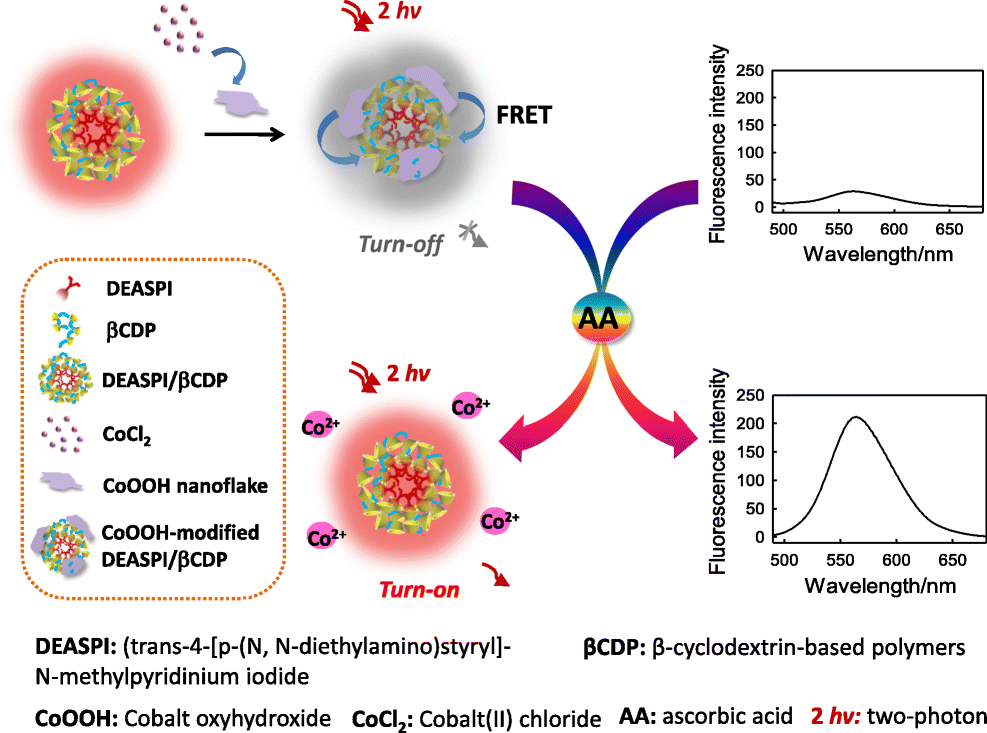
AbstractThis article describes the development of several nanoconjugates composed of cobalt (III) oxyhydroxide and DEASPI/βCDP, where DEASPI stands for the dye trans-4-[p-(N,N-diethylamino)styryl]-N-methylpyridinium, and βCDP stands for β-cyclodextrin. The material enables sensitive fluorometric detection and 3D imaging of ascorbic acid (AA) in biological samples. A nanomicelle composed of DEASPI and βCDP was prepared to act as a two-photon absorbance (TPA) nanofluorophore with desirable two-photon-sensitized fluorescence, high penetration depth, and excellent cell-permeability). The CoOOH nanoflakes were placed on the surface of the nanomicelle to act as both a quencher of fluorescence and as the recognition unit for AA. In the presence of AA, the CoOOH nanoflakes are reduced to Co (II), and this triggers the recovery of fluorescence. This new nanoprobe exhibits amplified two-photon fluorescence (excitation at 840 nm; emission at 565 nm), high sensitivity, and good selectivity. In-vitro imaging of endogenous AA was demonstrated in living HeLa cells. It was also employed to 3D imaging of exogenous AA in tissue by two-photon excitation microscopy to a depth of up to 320 μm. In our perception, this nanoprobe represents a valuable tool for elucidating the roles of AA in biochemical and clinical studies. Graphical abstractSchematic presentation of the preparation of a novel Poly β-Cyclodextrin/TPdye conjugated with cobalt oxyhydroxide nanoplatform and its application for high sensitive and two-photon 3D imaging of ascorbic acid (AA) in living cells and deep tissues.
中文翻译:

用聚-β-环糊精和花青染料修饰的羟基氧化钴作为活细胞和组织中抗坏血酸双光子成像的纳米平台
摘要本文描述了几种由羟基氧化钴 (III) 和 DEASPI/βCDP 组成的纳米复合物的开发,其中 DEASPI 代表染料 trans-4-[p-(N,N-diethylamino)styryl]-N-methylpyridinium,βCDP 代表染料β-环糊精。该材料能够对生物样品中的抗坏血酸 (AA) 进行灵敏的荧光检测和 3D 成像。制备由 DEASPI 和 βCDP 组成的纳米胶束作为双光子吸收 (TPA) 纳米荧光团,具有理想的双光子敏化荧光、高穿透深度和优异的细胞渗透性。CoOOH 纳米薄片被放置在纳米胶束的表面,作为荧光猝灭剂和 AA 的识别单元。在 AA 存在下,CoOOH 纳米薄片被还原为 Co (II),这触发了荧光的恢复。这种新型纳米探针具有放大的双光子荧光(840 nm 激发;565 nm 发射)、高灵敏度和良好的选择性。在活的 HeLa 细胞中证明了内源性 AA 的体外成像。它还被用于通过双光子激发显微镜对组织中的外源 AA 进行 3D 成像,深度可达 320 μm。在我们看来,这种纳米探针代表了阐明 AA 在生化和临床研究中的作用的宝贵工具。图形摘要与羟基氧化钴纳米平台共轭的新型聚 β-环糊精/TPdye 的制备及其在活细胞和深层组织中抗坏血酸 (AA) 的高灵敏度和双光子 3D 成像中的应用示意图。和良好的选择性。在活的 HeLa 细胞中证明了内源性 AA 的体外成像。它还被用于通过双光子激发显微镜对组织中的外源 AA 进行 3D 成像,深度可达 320 μm。在我们看来,这种纳米探针代表了阐明 AA 在生化和临床研究中的作用的宝贵工具。图形摘要与羟基氧化钴纳米平台共轭的新型聚 β-环糊精/TPdye 的制备及其在活细胞和深层组织中抗坏血酸 (AA) 的高灵敏度和双光子 3D 成像中的应用示意图。和良好的选择性。在活的 HeLa 细胞中证明了内源性 AA 的体外成像。它还被用于通过双光子激发显微镜对组织中的外源 AA 进行 3D 成像,深度可达 320 μm。在我们看来,这种纳米探针代表了阐明 AA 在生化和临床研究中的作用的宝贵工具。图形摘要与羟基氧化钴纳米平台共轭的新型聚 β-环糊精/TPdye 的制备及其在活细胞和深层组织中抗坏血酸 (AA) 的高灵敏度和双光子 3D 成像中的应用示意图。这种纳米探针是阐明 AA 在生化和临床研究中的作用的宝贵工具。图形摘要与羟基氧化钴纳米平台共轭的新型聚 β-环糊精/TPdye 的制备及其在活细胞和深层组织中抗坏血酸 (AA) 的高灵敏度和双光子 3D 成像中的应用示意图。这种纳米探针是阐明 AA 在生化和临床研究中的作用的宝贵工具。图形摘要与羟基氧化钴纳米平台共轭的新型聚 β-环糊精/TPdye 的制备及其在活细胞和深层组织中抗坏血酸 (AA) 的高灵敏度和双光子 3D 成像中的应用示意图。
更新日期:2019-02-22
中文翻译:

用聚-β-环糊精和花青染料修饰的羟基氧化钴作为活细胞和组织中抗坏血酸双光子成像的纳米平台
摘要本文描述了几种由羟基氧化钴 (III) 和 DEASPI/βCDP 组成的纳米复合物的开发,其中 DEASPI 代表染料 trans-4-[p-(N,N-diethylamino)styryl]-N-methylpyridinium,βCDP 代表染料β-环糊精。该材料能够对生物样品中的抗坏血酸 (AA) 进行灵敏的荧光检测和 3D 成像。制备由 DEASPI 和 βCDP 组成的纳米胶束作为双光子吸收 (TPA) 纳米荧光团,具有理想的双光子敏化荧光、高穿透深度和优异的细胞渗透性。CoOOH 纳米薄片被放置在纳米胶束的表面,作为荧光猝灭剂和 AA 的识别单元。在 AA 存在下,CoOOH 纳米薄片被还原为 Co (II),这触发了荧光的恢复。这种新型纳米探针具有放大的双光子荧光(840 nm 激发;565 nm 发射)、高灵敏度和良好的选择性。在活的 HeLa 细胞中证明了内源性 AA 的体外成像。它还被用于通过双光子激发显微镜对组织中的外源 AA 进行 3D 成像,深度可达 320 μm。在我们看来,这种纳米探针代表了阐明 AA 在生化和临床研究中的作用的宝贵工具。图形摘要与羟基氧化钴纳米平台共轭的新型聚 β-环糊精/TPdye 的制备及其在活细胞和深层组织中抗坏血酸 (AA) 的高灵敏度和双光子 3D 成像中的应用示意图。和良好的选择性。在活的 HeLa 细胞中证明了内源性 AA 的体外成像。它还被用于通过双光子激发显微镜对组织中的外源 AA 进行 3D 成像,深度可达 320 μm。在我们看来,这种纳米探针代表了阐明 AA 在生化和临床研究中的作用的宝贵工具。图形摘要与羟基氧化钴纳米平台共轭的新型聚 β-环糊精/TPdye 的制备及其在活细胞和深层组织中抗坏血酸 (AA) 的高灵敏度和双光子 3D 成像中的应用示意图。和良好的选择性。在活的 HeLa 细胞中证明了内源性 AA 的体外成像。它还被用于通过双光子激发显微镜对组织中的外源 AA 进行 3D 成像,深度可达 320 μm。在我们看来,这种纳米探针代表了阐明 AA 在生化和临床研究中的作用的宝贵工具。图形摘要与羟基氧化钴纳米平台共轭的新型聚 β-环糊精/TPdye 的制备及其在活细胞和深层组织中抗坏血酸 (AA) 的高灵敏度和双光子 3D 成像中的应用示意图。这种纳米探针是阐明 AA 在生化和临床研究中的作用的宝贵工具。图形摘要与羟基氧化钴纳米平台共轭的新型聚 β-环糊精/TPdye 的制备及其在活细胞和深层组织中抗坏血酸 (AA) 的高灵敏度和双光子 3D 成像中的应用示意图。这种纳米探针是阐明 AA 在生化和临床研究中的作用的宝贵工具。图形摘要与羟基氧化钴纳米平台共轭的新型聚 β-环糊精/TPdye 的制备及其在活细胞和深层组织中抗坏血酸 (AA) 的高灵敏度和双光子 3D 成像中的应用示意图。


 京公网安备 11010802027423号