当前位置:
X-MOL 学术
›
Microchim. Acta
›
论文详情
Our official English website, www.x-mol.net, welcomes your
feedback! (Note: you will need to create a separate account there.)
Raman spectroscopic imaging of pH values in cancerous tissue by using polyaniline@gold nanoparticles
Microchimica Acta ( IF 5.3 ) Pub Date : 2019-02-05 , DOI: 10.1007/s00604-019-3265-4 Zicheng Li , Ling Xia , Gongke Li , Yuling Hu
Microchimica Acta ( IF 5.3 ) Pub Date : 2019-02-05 , DOI: 10.1007/s00604-019-3265-4 Zicheng Li , Ling Xia , Gongke Li , Yuling Hu
|
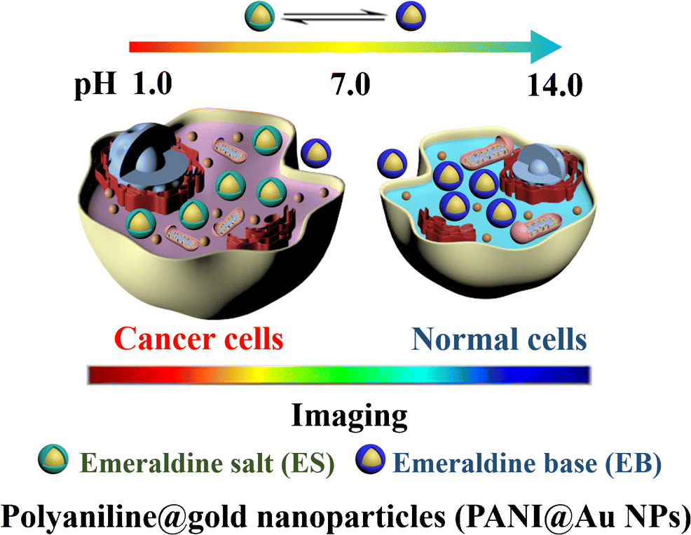
AbstractA core-shell nanocomposite consisting of polyaniline and gold nanoparticles (PANI@AuNPs) is shown to enable intracellular monitoring of pH values by surface-enhanced Raman scattering (SERS) spectroscopy. The method exploits the pH-responsive property of PANI and the SERS-enhancing effect of AuNPs. The intensity of the PANI Raman peak at 1164 cm−1 decreases on increasing the pH value from 4.6 to 7.4. This is the pH range encountered in normal cells and in cancer cells. The PANI@AuNPs were incorporated into HeLa cancer cells and 5 other kinds of cells for Raman based imaging of pH values. The results show that this pH nanoprobe can be applied for imaging of both normal cells and cancer cells. The core-shell composite was also applied to tissue imaging. In our perception, this core-shell nanoprobe is a valuable tool for imaging pH values of cancerous tissue. Graphical abstractSchematic presentation of a core-shell nanocomposite, polyaniline@gold nanoparticle, which was synthesized via a rapid method. With the pH of solution changing from alkaline to acidic, the polyaniline can change from emeraldine base (EB, blue shell) transition to emeraldine salt (ES, green shell) transition. Due to the pH-responsive property of polyaniline combined with the surface-enhanced Raman scattering spectroscopy effect of AuNPs. The polyaniline@gold nanoparticles were successfully applied as an intracellular pH probe.
中文翻译:

使用聚苯胺@金纳米粒子对癌组织中的 pH 值进行拉曼光谱成像
摘要 由聚苯胺和金纳米粒子 (PANI@AuNPs) 组成的核壳纳米复合材料可以通过表面增强拉曼散射 (SERS) 光谱法对 pH 值进行细胞内监测。该方法利用了 PANI 的 pH 响应特性和 AuNP 的 SERS 增强效应。随着 pH 值从 4.6 增加到 7.4,1164 cm-1 处的 PANI 拉曼峰强度降低。这是正常细胞和癌细胞中遇到的 pH 值范围。PANI@AuNPs 被整合到 HeLa 癌细胞和其他 5 种细胞中,用于基于拉曼的 pH 值成像。结果表明,这种 pH 纳米探针可用于正常细胞和癌细胞的成像。核壳复合材料也应用于组织成像。在我们的认知中,这种核壳纳米探针是对癌组织的 pH 值进行成像的宝贵工具。图形摘要通过快速方法合成的核壳纳米复合材料聚苯胺@金纳米颗粒的示意图。随着溶液的pH值由碱性变为酸性,聚苯胺可以从翡翠碱(EB,蓝壳)转变为翡翠盐(ES,绿壳)转变。由于聚苯胺的 pH 响应特性与 AuNP 的表面增强拉曼散射光谱效应相结合。聚苯胺@金纳米粒子被成功地用作细胞内 pH 探针。聚苯胺可以从翡翠碱(EB,蓝壳)过渡到翡翠盐(ES,绿壳)过渡。由于聚苯胺的 pH 响应特性与 AuNP 的表面增强拉曼散射光谱效应相结合。聚苯胺@金纳米粒子被成功地用作细胞内 pH 探针。聚苯胺可以从翡翠碱(EB,蓝壳)过渡到翡翠盐(ES,绿壳)过渡。由于聚苯胺的 pH 响应特性与 AuNP 的表面增强拉曼散射光谱效应相结合。聚苯胺@金纳米粒子被成功地用作细胞内 pH 探针。
更新日期:2019-02-05
中文翻译:
使用聚苯胺@金纳米粒子对癌组织中的 pH 值进行拉曼光谱成像
摘要 由聚苯胺和金纳米粒子 (PANI@AuNPs) 组成的核壳纳米复合材料可以通过表面增强拉曼散射 (SERS) 光谱法对 pH 值进行细胞内监测。该方法利用了 PANI 的 pH 响应特性和 AuNP 的 SERS 增强效应。随着 pH 值从 4.6 增加到 7.4,1164 cm-1 处的 PANI 拉曼峰强度降低。这是正常细胞和癌细胞中遇到的 pH 值范围。PANI@AuNPs 被整合到 HeLa 癌细胞和其他 5 种细胞中,用于基于拉曼的 pH 值成像。结果表明,这种 pH 纳米探针可用于正常细胞和癌细胞的成像。核壳复合材料也应用于组织成像。在我们的认知中,这种核壳纳米探针是对癌组织的 pH 值进行成像的宝贵工具。图形摘要通过快速方法合成的核壳纳米复合材料聚苯胺@金纳米颗粒的示意图。随着溶液的pH值由碱性变为酸性,聚苯胺可以从翡翠碱(EB,蓝壳)转变为翡翠盐(ES,绿壳)转变。由于聚苯胺的 pH 响应特性与 AuNP 的表面增强拉曼散射光谱效应相结合。聚苯胺@金纳米粒子被成功地用作细胞内 pH 探针。聚苯胺可以从翡翠碱(EB,蓝壳)过渡到翡翠盐(ES,绿壳)过渡。由于聚苯胺的 pH 响应特性与 AuNP 的表面增强拉曼散射光谱效应相结合。聚苯胺@金纳米粒子被成功地用作细胞内 pH 探针。聚苯胺可以从翡翠碱(EB,蓝壳)过渡到翡翠盐(ES,绿壳)过渡。由于聚苯胺的 pH 响应特性与 AuNP 的表面增强拉曼散射光谱效应相结合。聚苯胺@金纳米粒子被成功地用作细胞内 pH 探针。


 京公网安备 11010802027423号