Spectrochimica Acta Part A: Molecular and Biomolecular Spectroscopy ( IF 4.3 ) Pub Date : 2019-02-05 , DOI: 10.1016/j.saa.2019.02.006 Andrés Vásquez-Rivera , Harriëtte Oldenhof , Andres Hilfiker , Willem F. Wolkers
|
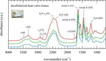
Decellularized heart valves hold promise for their use as bioscaffolds in cardiovascular surgery. Quality assessment of heart valves after decellularization processing and/or storage is time consuming and destructive. Fourier transform infrared spectroscopy (FTIR) allows rapid non-invasive assessment of biomolecular structures in tissues. In this study, IR-spectra taken from different layers of the pulmonary artery trunk and leaflet tissues of decellularized porcine heart valves were compared with those of pure collagen and elastin, the main protein components in these tissues. In addition, spectral changes associated with aging and oxidative damage were investigated. Infrared absorbance spectra of the arteria intima and media layer were found to be very similar, whereas distinct differences were observed when compared with spectra of the externa layer. In the latter, the shape of the CH-stretching vibration region (3050–2800 cm−1) resembled that of pure collagen. Also, pronounced νCOOH and amide-II bands and a relatively high content of α-helical structures in the externa layer indicated the presence of collagen in this layer. The externa layer of the artery appeared to be sensitive to collagenase treatment, whereas the media and intima layer were particularly affected by elastase and not by collagenase treatment. Protein conformational changes after treatment with collagenase were observed in all three layers. Collagenase treatment completely degraded the leaflet tissue sections. Spectra were also collected from scaffolds after 2 and 12 weeks storage at 37 °C, and after induced oxidative damage. Spectral changes related to aging and oxidative damage were particularly evident in the CH-stretching region, whereas the shape of the amide-I band, reflecting the overall protein secondary structure, remained unaltered.
中文翻译:
去细胞心脏瓣膜支架的光谱指纹图谱
去细胞心脏瓣膜有望在心血管外科手术中用作生物支架。脱细胞处理和/或储存后心脏瓣膜的质量评估既费时又具有破坏性。傅里叶变换红外光谱(FTIR)可以对组织中的生物分子结构进行快速的非侵入性评估。在这项研究中,比较了从脱细胞的猪心脏瓣膜的肺动脉主干和小叶组织的不同层获取的红外光谱与纯胶原蛋白和弹性蛋白(这些组织中的主要蛋白质成分)的红外光谱。此外,还研究了与老化和氧化损伤相关的光谱变化。发现动脉内膜和培养基层的红外吸收光谱非常相似,与外部层的光谱相比,观察到了明显的差异。在后者中,CH拉伸振动区域的形状(3050–2800 cm-1)类似于纯胶原蛋白。同样,外部层中明显的νCOOH和酰胺-II带以及相对较高的α-螺旋结构含量表明该层中存在胶原蛋白。动脉的外层似乎对胶原酶治疗敏感,而中层和内膜层特别受弹性蛋白酶而不是胶原酶治疗的影响。在所有三个层中均观察到用胶原酶处理后的蛋白质构象变化。胶原酶处理完全降解了小叶组织切片。在37°C下保存2周和12周以及诱导的氧化损伤后,还从支架中收集光谱。与衰老和氧化损伤有关的光谱变化在CH拉伸区域尤为明显,而酰胺I谱带的形状


 京公网安备 11010802027423号