当前位置:
X-MOL 学术
›
Adv. Mater.
›
论文详情
Our official English website, www.x-mol.net, welcomes your
feedback! (Note: you will need to create a separate account there.)
Real‐Time and High‐Resolution Bioimaging with Bright Aggregation‐Induced Emission Dots in Short‐Wave Infrared Region
Advanced Materials ( IF 27.4 ) Pub Date : 2018-01-17 , DOI: 10.1002/adma.201706856 Ji Qi 1 , Chaowei Sun 2 , Abudureheman Zebibula 3 , Hequn Zhang 2 , Ryan T. K. Kwok 1 , Xinyuan Zhao 4 , Wang Xi 5 , Jacky W. Y. Lam 1 , Jun Qian 2 , Ben Zhong Tang 1
Advanced Materials ( IF 27.4 ) Pub Date : 2018-01-17 , DOI: 10.1002/adma.201706856 Ji Qi 1 , Chaowei Sun 2 , Abudureheman Zebibula 3 , Hequn Zhang 2 , Ryan T. K. Kwok 1 , Xinyuan Zhao 4 , Wang Xi 5 , Jacky W. Y. Lam 1 , Jun Qian 2 , Ben Zhong Tang 1
Affiliation

|
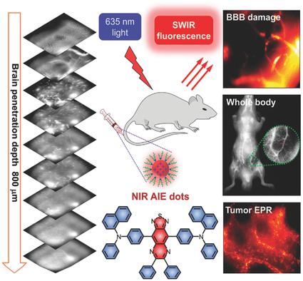
Fluorescence imaging in the spectral region beyond the conventional near‐infrared biological window (700–900 nm) can theoretically afford high resolution and deep tissue penetration. Although some efforts have been devoted to developing a short‐wave infrared (SWIR; 900–1700 nm) imaging modality in the past decade, long‐wavelength biomedical imaging is still suboptimal owing to the unsatisfactory materials properties of SWIR fluorophores. Taking advantage of organic dots based on an aggregation‐induced emission luminogen (AIEgen), herein microscopic vasculature imaging of brain and tumor is reported in living mice in the SWIR spectral region. The long‐wavelength emission of AIE dots with certain brightness facilitates resolving brain capillaries with high spatial resolution (≈3 µm) and deep penetration (800 µm). Owning to the deep penetration depth and real‐time imaging capability, in vivo SWIR microscopic angiography exhibits superior resolution in monitoring blood–brain barrier damage in mouse brain, and visualizing enhanced permeability and retention effect in tumor sites. Furthermore, the AIE dots show good biocompatibility, and no noticeable abnormalities, inflammations or lesions are observed in the main organs of the mice. This work will inspire new insights on development of advanced SWIR techniques for biomedical imaging.
中文翻译:

短波红外区域中具有明亮聚集诱导发射点的实时和高分辨率生物成像
从理论上讲,在常规近红外生物窗口(700-900 nm)以外的光谱区域进行荧光成像可以提供高分辨率和深层组织穿透能力。尽管在过去十年中已经致力于开发短波红外(SWIR; 900-1700 nm)成像方式,但是由于SWIR荧光团的材料性能不理想,长波生物医学成像仍然不是最佳选择。利用基于聚集诱导的发光发光剂(AIEgen)的有机点,据报道在活体小鼠的SWIR光谱区域中,大脑和肿瘤的显微脉管系统成像得到了报道。具有一定亮度的AIE点的长波长发射有助于解析具有高空间分辨率(≈3µm)和深度穿透(800 µm)的脑毛细血管。凭借深层的穿透深度和实时成像功能,体内SWIR显微血管造影在监测小鼠脑血脑屏障损害以及可视化增强的肿瘤通透性和保留位点效果方面显示出卓越的分辨率。此外,AIE点显示出良好的生物相容性,并且在小鼠的主要器官中未观察到明显的异常,发炎或病变。这项工作将激发有关生物医学成像高级SWIR技术开发的新见识。在小鼠的主要器官中观察到炎症或损伤。这项工作将激发有关生物医学成像高级SWIR技术开发的新见解。在小鼠的主要器官中观察到炎症或损伤。这项工作将激发有关生物医学成像高级SWIR技术开发的新见识。
更新日期:2018-01-17
中文翻译:

短波红外区域中具有明亮聚集诱导发射点的实时和高分辨率生物成像
从理论上讲,在常规近红外生物窗口(700-900 nm)以外的光谱区域进行荧光成像可以提供高分辨率和深层组织穿透能力。尽管在过去十年中已经致力于开发短波红外(SWIR; 900-1700 nm)成像方式,但是由于SWIR荧光团的材料性能不理想,长波生物医学成像仍然不是最佳选择。利用基于聚集诱导的发光发光剂(AIEgen)的有机点,据报道在活体小鼠的SWIR光谱区域中,大脑和肿瘤的显微脉管系统成像得到了报道。具有一定亮度的AIE点的长波长发射有助于解析具有高空间分辨率(≈3µm)和深度穿透(800 µm)的脑毛细血管。凭借深层的穿透深度和实时成像功能,体内SWIR显微血管造影在监测小鼠脑血脑屏障损害以及可视化增强的肿瘤通透性和保留位点效果方面显示出卓越的分辨率。此外,AIE点显示出良好的生物相容性,并且在小鼠的主要器官中未观察到明显的异常,发炎或病变。这项工作将激发有关生物医学成像高级SWIR技术开发的新见识。在小鼠的主要器官中观察到炎症或损伤。这项工作将激发有关生物医学成像高级SWIR技术开发的新见解。在小鼠的主要器官中观察到炎症或损伤。这项工作将激发有关生物医学成像高级SWIR技术开发的新见识。


 京公网安备 11010802027423号