当前位置:
X-MOL 学术
›
Biotechnol. J.
›
论文详情
Our official English website, www.x-mol.net, welcomes your
feedback! (Note: you will need to create a separate account there.)
“Cyt/Nuc,” a Customizable and Documenting ImageJ Macro for Evaluation of Protein Distributions Between Cytosol and Nucleus
Biotechnology Journal ( IF 3.2 ) Pub Date : 2018-02-09 , DOI: 10.1002/biot.201700652 Tilman Grune 1, 2, 3, 4 , Richard Kehm 1, 2 , Annika Höhn 1, 2 , Tobias Jung 1, 3
Biotechnology Journal ( IF 3.2 ) Pub Date : 2018-02-09 , DOI: 10.1002/biot.201700652 Tilman Grune 1, 2, 3, 4 , Richard Kehm 1, 2 , Annika Höhn 1, 2 , Tobias Jung 1, 3
Affiliation
|
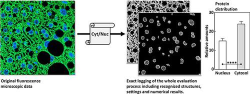
Large amounts of data from multi‐channel, high resolution, fluorescence microscopic images require tools that provide easy, customizable, and reproducible high‐throughput analysis. The freeware “ImageJ” has become one of the standard tools for scientific image analysis. Since ImageJ offers recording of “macros,” even a complex multi‐step process can be easily applied fully automated to large numbers of images, saving both time and reducing human subjective evaluation. In this work, we present “Cyt/Nuc,” an ImageJ macro, able to recognize and to compare the nuclear and cytosolic areas of tissue samples, in order to investigate distributions of immunostained proteins between both compartments, while it documents in detail the whole process of evaluation and pattern recognition. As practical example, the redistribution of the 20S proteasome, the main intracellular protease in mammalian cells, is investigated in NZO‐mouse liver after feeding the animals different diets. A significant shift in proteasomal distribution between cytosol and nucleus in response to metabolic stress was revealed using “Cyt/Nuc” via automatized quantification of thousands of nuclei within minutes. “Cyt/Nuc” is easy to use and highly customizable, matches the precision of careful manual evaluation and bears the potential for quick detection of any shift in intracellular protein distribution.
中文翻译:

“ Cyt / Nuc”,一个可自定义和记录文档的ImageJ宏,用于评估细胞溶胶和细胞核之间的蛋白质分布
来自多通道,高分辨率,荧光显微图像的大量数据需要工具来提供容易,可定制和可重现的高通量分析。免费软件“ ImageJ”已成为用于科学图像分析的标准工具之一。由于ImageJ提供了“宏”的记录,因此即使是复杂的多步骤过程,也可以轻松地将其自动化地自动应用于大量图像,从而节省了时间并减少了人的主观评估。在这项工作中,我们展示了ImageJ宏“ Cyt / Nuc”,它能够识别并比较组织样本的核和胞质区域,以便研究免疫染色的蛋白在两个区室之间的分布,同时详细记录了整个过程评估和模式识别的过程。举一个实际的例子,20S蛋白酶体的重新分布,喂养动物不同饮食后,在NZO小鼠肝脏中研究了哺乳动物细胞中的主要细胞内蛋白酶。通过使用“ Cyt / Nuc”,通过在几分钟内对数千个核的自动定量,揭示了响应代谢应激,胞质和核之间的蛋白酶体分布发生了显着变化。“ Cyt / Nuc”易于使用且可高度自定义,具有经过仔细人工评估的精度,并具有快速检测细胞内蛋白质分布变化的潜力。通过使用“ Cyt / Nuc”,通过在几分钟内对数千个核的自动定量,揭示了响应代谢应激,胞质和核之间的蛋白酶体分布发生了显着变化。“ Cyt / Nuc”易于使用且可高度自定义,具有经过仔细人工评估的精度,并具有快速检测细胞内蛋白质分布变化的潜力。通过使用“ Cyt / Nuc”,通过在几分钟内对数千个核的自动定量,揭示了响应代谢应激,胞质和核之间的蛋白酶体分布发生了显着变化。“ Cyt / Nuc”易于使用且可高度自定义,具有经过仔细人工评估的精度,并具有快速检测细胞内蛋白质分布变化的潜力。
更新日期:2018-03-21
中文翻译:
“ Cyt / Nuc”,一个可自定义和记录文档的ImageJ宏,用于评估细胞溶胶和细胞核之间的蛋白质分布
来自多通道,高分辨率,荧光显微图像的大量数据需要工具来提供容易,可定制和可重现的高通量分析。免费软件“ ImageJ”已成为用于科学图像分析的标准工具之一。由于ImageJ提供了“宏”的记录,因此即使是复杂的多步骤过程,也可以轻松地将其自动化地自动应用于大量图像,从而节省了时间并减少了人的主观评估。在这项工作中,我们展示了ImageJ宏“ Cyt / Nuc”,它能够识别并比较组织样本的核和胞质区域,以便研究免疫染色的蛋白在两个区室之间的分布,同时详细记录了整个过程评估和模式识别的过程。举一个实际的例子,20S蛋白酶体的重新分布,喂养动物不同饮食后,在NZO小鼠肝脏中研究了哺乳动物细胞中的主要细胞内蛋白酶。通过使用“ Cyt / Nuc”,通过在几分钟内对数千个核的自动定量,揭示了响应代谢应激,胞质和核之间的蛋白酶体分布发生了显着变化。“ Cyt / Nuc”易于使用且可高度自定义,具有经过仔细人工评估的精度,并具有快速检测细胞内蛋白质分布变化的潜力。通过使用“ Cyt / Nuc”,通过在几分钟内对数千个核的自动定量,揭示了响应代谢应激,胞质和核之间的蛋白酶体分布发生了显着变化。“ Cyt / Nuc”易于使用且可高度自定义,具有经过仔细人工评估的精度,并具有快速检测细胞内蛋白质分布变化的潜力。通过使用“ Cyt / Nuc”,通过在几分钟内对数千个核的自动定量,揭示了响应代谢应激,胞质和核之间的蛋白酶体分布发生了显着变化。“ Cyt / Nuc”易于使用且可高度自定义,具有经过仔细人工评估的精度,并具有快速检测细胞内蛋白质分布变化的潜力。


 京公网安备 11010802027423号