当前位置:
X-MOL 学术
›
Neuropsychologia
›
论文详情
Our official English website, www.x-mol.net, welcomes your
feedback! (Note: you will need to create a separate account there.)
Subcortical pathways to extrastriate visual cortex underlie residual vision following bilateral damage to V1.
Neuropsychologia ( IF 2.0 ) Pub Date : 2018-01-07 , DOI: 10.1016/j.neuropsychologia.2018.01.007 Sara Ajina 1 , Holly Bridge 1
Neuropsychologia ( IF 2.0 ) Pub Date : 2018-01-07 , DOI: 10.1016/j.neuropsychologia.2018.01.007 Sara Ajina 1 , Holly Bridge 1
Affiliation
|
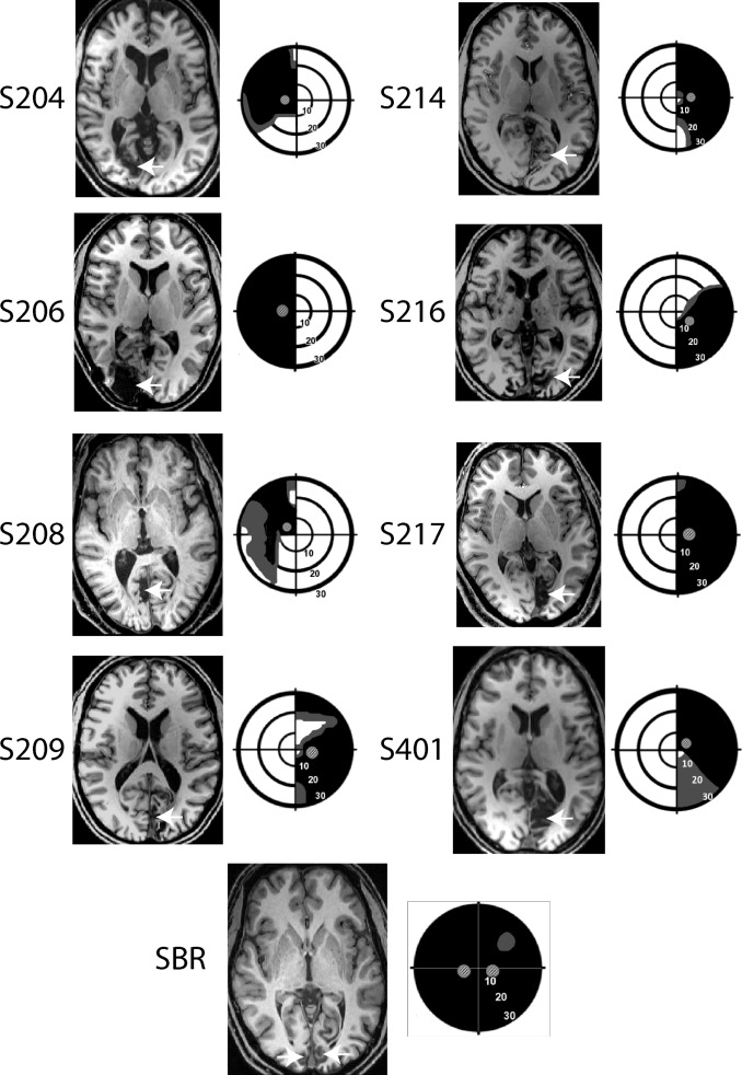
Residual vision, or blindsight, following damage to the primary visual cortex (V1) has been investigated for almost half a century. While there have been many studies of patients with unilateral damage to V1, far fewer have examined bilateral damage, mainly due to the rarity of such patients. Here we re-examine the residual visual function and underlying pathways of previously studied patient SBR who, as a young adult, suffered bilateral damage restricted to V1 which rendered him cortically blind. While earlier work compared his visual cortex to healthy, sighted participants, here we consider how his visual responses and connections compare to patients with unilateral damage to V1 in addition to sighted participants. Detection of drifting Gabor patches of different contrasts (1%, 5%, 10%, 50% and 100%) was tested in SBR and a group of eight patients with unilateral damage to V1. Performance was compared to the neural activation in motion area hMT+ measured using functional magnetic resonance imaging. Diffusion tractography was also used to determine the white matter microstructure of the visual pathways in all participants. Like the patients with unilateral damage, patient SBR showed increased % BOLD signal change to the high contrast stimuli that he could detect compared to the lower contrast stimuli that were not detectable. Diffusion tractography suggests this information is conveyed by a direct pathway between the lateral geniculate nucleus (LGN) and hMT+ since this pathway had microstructure that was comparable to the healthy control group. In contrast, the pathway between LGN and V1 had reduced integrity compared to controls. A further finding of note was that, unlike control participants, SBR showed similar patterns of contralateral and ipsilateral activity in hMT+, in addition to healthy white matter microstructure in the tract connecting hMT+ between the two hemispheres. This raises the possibility of increased connectivity between the two hemispheres in the absence of V1 input. In conclusion, the pattern of visual function and anatomy in bilateral cortical damage is comparable to that seen in a group of patients with unilateral damage. Thus, while the intact hemisphere may play a role in residual vision in patients with unilateral damage, its influence is not evident with the methodology employed here.
中文翻译:

在 V1 双侧损伤后,皮层外视皮层的皮层下通路是残余视力的基础。
近半个世纪以来,人们已经研究了初级视觉皮层 (V1) 受损后的残余视力或盲视。虽然有许多关于 V1 单侧损伤患者的研究,但很少有研究双侧损伤,主要是因为此类患者很少见。在这里,我们重新检查了先前研究的患者 SBR 的残余视觉功能和潜在通路,他们作为一个年轻的成年人,遭受了仅限于 V1 的双侧损伤,导致他的皮质失明。虽然早期的工作将他的视觉皮层与健康、有视力的参与者进行了比较,但在这里我们考虑了他的视觉反应和连接如何与除了有视力的参与者之外的 V1 单侧损伤的患者进行比较。检测不同对比度(1%、5%、10%、50% 和 100%)在 SBR 和一组 8 名单侧 V1 损伤患者中进行了测试。将性能与使用功能性磁共振成像测量的运动区域 hMT+ 中的神经激活进行比较。扩散束成像也用于确定所有参与者视觉通路的白质微结构。与具有单侧损伤的患者一样,与无法检测到的较低对比度刺激相比,患者 SBR 对他可以检测到的高对比度刺激显示增加的 % BOLD 信号变化。扩散束成像表明该信息通过外侧膝状体核 (LGN) 和 hMT+ 之间的直接通路传递,因为该通路具有与健康对照组相当的微观结构。相反,与对照组相比,LGN 和 V1 之间的通路完整性降低。另一个值得注意的发现是,与对照组参与者不同,SBR 在 hMT+ 中显示出相似的对侧和同侧活动模式,除了连接两个半球之间的 hMT+ 的通道中的健康白质微结构。这增加了在没有 V1 输入的情况下增加两个半球之间连接的可能性。总之,双侧皮质损伤的视觉功能和解剖模式与一组单侧损伤患者的相似。因此,虽然完整的半球可能在单侧损伤患者的残余视力中发挥作用,但其影响在此处采用的方法中并不明显。除了连接两个半球之间的 hMT+ 的管道中的健康白质微结构。这增加了在没有 V1 输入的情况下增加两个半球之间连接的可能性。总之,双侧皮质损伤的视觉功能和解剖模式与一组单侧损伤患者的相似。因此,虽然完整的半球可能在单侧损伤患者的残余视力中发挥作用,但其影响在此处采用的方法中并不明显。除了连接两个半球之间的 hMT+ 的管道中的健康白质微结构。这增加了在没有 V1 输入的情况下增加两个半球之间连接的可能性。总之,双侧皮质损伤的视觉功能和解剖模式与一组单侧损伤患者的相似。因此,虽然完整的半球可能在单侧损伤患者的残余视力中发挥作用,但其影响在此处采用的方法中并不明显。双侧皮质损伤的视觉功能和解剖模式与一组单侧损伤患者的情况相当。因此,虽然完整的半球可能在单侧损伤患者的残余视力中发挥作用,但其影响在此处采用的方法中并不明显。双侧皮质损伤的视觉功能和解剖模式与一组单侧损伤患者的情况相当。因此,虽然完整的半球可能在单侧损伤患者的残余视力中发挥作用,但其影响在此处采用的方法中并不明显。
更新日期:2018-01-07
中文翻译:
在 V1 双侧损伤后,皮层外视皮层的皮层下通路是残余视力的基础。
近半个世纪以来,人们已经研究了初级视觉皮层 (V1) 受损后的残余视力或盲视。虽然有许多关于 V1 单侧损伤患者的研究,但很少有研究双侧损伤,主要是因为此类患者很少见。在这里,我们重新检查了先前研究的患者 SBR 的残余视觉功能和潜在通路,他们作为一个年轻的成年人,遭受了仅限于 V1 的双侧损伤,导致他的皮质失明。虽然早期的工作将他的视觉皮层与健康、有视力的参与者进行了比较,但在这里我们考虑了他的视觉反应和连接如何与除了有视力的参与者之外的 V1 单侧损伤的患者进行比较。检测不同对比度(1%、5%、10%、50% 和 100%)在 SBR 和一组 8 名单侧 V1 损伤患者中进行了测试。将性能与使用功能性磁共振成像测量的运动区域 hMT+ 中的神经激活进行比较。扩散束成像也用于确定所有参与者视觉通路的白质微结构。与具有单侧损伤的患者一样,与无法检测到的较低对比度刺激相比,患者 SBR 对他可以检测到的高对比度刺激显示增加的 % BOLD 信号变化。扩散束成像表明该信息通过外侧膝状体核 (LGN) 和 hMT+ 之间的直接通路传递,因为该通路具有与健康对照组相当的微观结构。相反,与对照组相比,LGN 和 V1 之间的通路完整性降低。另一个值得注意的发现是,与对照组参与者不同,SBR 在 hMT+ 中显示出相似的对侧和同侧活动模式,除了连接两个半球之间的 hMT+ 的通道中的健康白质微结构。这增加了在没有 V1 输入的情况下增加两个半球之间连接的可能性。总之,双侧皮质损伤的视觉功能和解剖模式与一组单侧损伤患者的相似。因此,虽然完整的半球可能在单侧损伤患者的残余视力中发挥作用,但其影响在此处采用的方法中并不明显。除了连接两个半球之间的 hMT+ 的管道中的健康白质微结构。这增加了在没有 V1 输入的情况下增加两个半球之间连接的可能性。总之,双侧皮质损伤的视觉功能和解剖模式与一组单侧损伤患者的相似。因此,虽然完整的半球可能在单侧损伤患者的残余视力中发挥作用,但其影响在此处采用的方法中并不明显。除了连接两个半球之间的 hMT+ 的管道中的健康白质微结构。这增加了在没有 V1 输入的情况下增加两个半球之间连接的可能性。总之,双侧皮质损伤的视觉功能和解剖模式与一组单侧损伤患者的相似。因此,虽然完整的半球可能在单侧损伤患者的残余视力中发挥作用,但其影响在此处采用的方法中并不明显。双侧皮质损伤的视觉功能和解剖模式与一组单侧损伤患者的情况相当。因此,虽然完整的半球可能在单侧损伤患者的残余视力中发挥作用,但其影响在此处采用的方法中并不明显。双侧皮质损伤的视觉功能和解剖模式与一组单侧损伤患者的情况相当。因此,虽然完整的半球可能在单侧损伤患者的残余视力中发挥作用,但其影响在此处采用的方法中并不明显。


 京公网安备 11010802027423号