Our official English website, www.x-mol.net, welcomes your
feedback! (Note: you will need to create a separate account there.)
Molecular Mechanism and Dynamics of S-Deoxyephedrine Moving through Molecular Channels within D3R
ACS Omega ( IF 3.7 ) Pub Date : 2017-12-13 00:00:00 , DOI: 10.1021/acsomega.7b01161 Ai Jing Li 1 , Wei Xie 1 , Ming Wang 1 , Si Chuan Xu 1
ACS Omega ( IF 3.7 ) Pub Date : 2017-12-13 00:00:00 , DOI: 10.1021/acsomega.7b01161 Ai Jing Li 1 , Wei Xie 1 , Ming Wang 1 , Si Chuan Xu 1
Affiliation

|
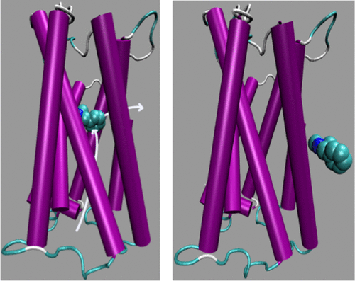
In this article, the trajectories of S-deoxyephedrine (SBD) along molecular channels within the complex protein structure of third dopamine receptor (D3R) are analyzed via molecular dynamic techniques, including potential mean force calculations of umbrella samplings from the 4.5 version of the GROMACS program. Changes in free energy due to the movement of SBD within D3R are determined, and the molecular dynamic mechanisms of SBD transmitting along molecular channels are probed. Molecular simulated results show that the change in free energy is calculated as 171.7 kJ·mol–1 for the transmission of SBD toward the outside of the cell along the y+ axis functional molecular channel and is 275.0 kJ·mol–1 for movement toward the intracellular structure along the y– axis. Within the internal structure of D3R, the changes in free energy are determined to be 103.6, 242.1, 459.7, and 127.8 kJ·mol–1 for transmission of SBD along the x+, x–, z+, and z– axes, respectively, toward the cell bilayer membrane, which indicates that SBD leaves much more easily along the x+ axis through the gap between the TM5 (the fifth transmembrane helix) and TM6 (the sixth transmembrane helix) from the internal structure of D3R. The values of free-energy changes indicate that SBD molecules can clear the protective channel within D3R, which helps dopamine molecules to leave the D3R internal structure along the x+ axis and to prevent them for exerting excessive neurotransmitter function. Therefore, our results suggest that SBD is effective for development as a drug for treating schizophrenia and its pharmacology is closely related to its dynamics and mechanisms within the molecular pathway of dopamine receptors.
中文翻译:

S-脱氧麻黄碱通过D 3 R内分子通道的分子机理和动力学
在本文中,通过分子动力学技术分析了第三多巴胺受体(D 3 R)复杂蛋白质结构内沿S-脱氧麻黄碱(SBD)的分子通道的轨迹,包括从4.5版本的伞状样品中潜在的平均力计算GROMACS程序。确定了由于SBD在D 3 R中的运动而引起的自由能变化,并探究了SBD沿分子通道传输的分子动力学机理。分子模拟结果表明,SBD沿y +轴功能分子通道向细胞外部的传输自由能变化为171.7 kJ·mol –1,为275.0 kJ·mol –1–1用于沿y轴向细胞内结构移动。在D 3 R的内部结构中,对于SBD沿x +,x –,z +和z –轴的传输,自由能的变化确定为103.6、242.1、459.7和127.8 kJ·mol –1。分别朝向细胞双层膜,这表明SBD从D 3的内部结构更容易沿着x +轴通过TM5(第五个跨膜螺旋)和TM6(第六个跨膜螺旋)之间的间隙离开。R.自由能变化的值表明SBD分子可以清除D 3 R中的保护通道,这有助于多巴胺分子沿x +轴离开D 3 R内部结构,并防止它们发挥过度的神经递质功能。因此,我们的结果表明,SBD作为治疗精神分裂症的药物可有效发展,其药理作用与其在多巴胺受体分子途径内的动力学和机制密切相关。
更新日期:2017-12-13
中文翻译:

S-脱氧麻黄碱通过D 3 R内分子通道的分子机理和动力学
在本文中,通过分子动力学技术分析了第三多巴胺受体(D 3 R)复杂蛋白质结构内沿S-脱氧麻黄碱(SBD)的分子通道的轨迹,包括从4.5版本的伞状样品中潜在的平均力计算GROMACS程序。确定了由于SBD在D 3 R中的运动而引起的自由能变化,并探究了SBD沿分子通道传输的分子动力学机理。分子模拟结果表明,SBD沿y +轴功能分子通道向细胞外部的传输自由能变化为171.7 kJ·mol –1,为275.0 kJ·mol –1–1用于沿y轴向细胞内结构移动。在D 3 R的内部结构中,对于SBD沿x +,x –,z +和z –轴的传输,自由能的变化确定为103.6、242.1、459.7和127.8 kJ·mol –1。分别朝向细胞双层膜,这表明SBD从D 3的内部结构更容易沿着x +轴通过TM5(第五个跨膜螺旋)和TM6(第六个跨膜螺旋)之间的间隙离开。R.自由能变化的值表明SBD分子可以清除D 3 R中的保护通道,这有助于多巴胺分子沿x +轴离开D 3 R内部结构,并防止它们发挥过度的神经递质功能。因此,我们的结果表明,SBD作为治疗精神分裂症的药物可有效发展,其药理作用与其在多巴胺受体分子途径内的动力学和机制密切相关。


 京公网安备 11010802027423号