Our official English website, www.x-mol.net, welcomes your
feedback! (Note: you will need to create a separate account there.)
Three-Dimensional Reconstruction of Nuclear Envelope Architecture Using Dual-Color Metal-Induced Energy Transfer Imaging
ACS Nano ( IF 15.8 ) Pub Date : 2017-09-20 00:00:00 , DOI: 10.1021/acsnano.7b04671 Anna M. Chizhik 1 , Daja Ruhlandt 1 , Janine Pfaff 2 , Narain Karedla 1 , Alexey I. Chizhik 1 , Ingo Gregor 1 , Ralph H. Kehlenbach 2 , Jörg Enderlein 1
ACS Nano ( IF 15.8 ) Pub Date : 2017-09-20 00:00:00 , DOI: 10.1021/acsnano.7b04671 Anna M. Chizhik 1 , Daja Ruhlandt 1 , Janine Pfaff 2 , Narain Karedla 1 , Alexey I. Chizhik 1 , Ingo Gregor 1 , Ralph H. Kehlenbach 2 , Jörg Enderlein 1
Affiliation

|
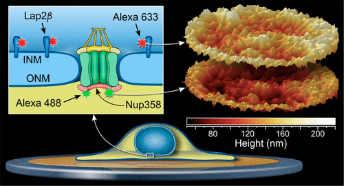
The nuclear envelope, comprising the inner and the outer nuclear membrane, separates the nucleus from the cytoplasm and plays a key role in cellular functions. Nuclear pore complexes (NPCs), which are embedded in the nuclear envelope, control transport of macromolecules between the two compartments. Here, using dual-color metal-induced energy transfer (MIET), we determine the axial distance between Lap2β and Nup358 as markers for the inner nuclear membrane and the cytoplasmic side of the NPC, respectively. Using MIET imaging, we reconstruct the 3D profile of the nuclear envelope over the whole basal area, with an axial resolution of a few nanometers. This result demonstrates that optical microscopy can achieve nanometer axial resolution in biological samples and without recourse to complex interferometric approaches.
中文翻译:

使用双色金属诱导的能量转移成像三维重建核信封结构
包含内部和外部核膜的核被膜将细胞核与细胞质分开,并在细胞功能中发挥关键作用。嵌入核膜中的核孔复合物(NPC)控制大分子在两个隔室之间的运输。在这里,我们使用双色金属诱导的能量转移(MIET),确定Lap2β和Nup358之间的轴向距离分别作为内核膜和NPC胞质侧的标记。使用MIET成像,我们可以在整个基础区域上重建核包膜的3D轮廓,其轴向分辨率为几纳米。该结果表明,光学显微镜可以在生物样品中实现纳米轴向分辨率,而无需求助于复杂的干涉测量方法。
更新日期:2017-09-20
中文翻译:

使用双色金属诱导的能量转移成像三维重建核信封结构
包含内部和外部核膜的核被膜将细胞核与细胞质分开,并在细胞功能中发挥关键作用。嵌入核膜中的核孔复合物(NPC)控制大分子在两个隔室之间的运输。在这里,我们使用双色金属诱导的能量转移(MIET),确定Lap2β和Nup358之间的轴向距离分别作为内核膜和NPC胞质侧的标记。使用MIET成像,我们可以在整个基础区域上重建核包膜的3D轮廓,其轴向分辨率为几纳米。该结果表明,光学显微镜可以在生物样品中实现纳米轴向分辨率,而无需求助于复杂的干涉测量方法。


 京公网安备 11010802027423号