当前位置:
X-MOL 学术
›
ACS Biomater. Sci. Eng.
›
论文详情
Our official English website, www.x-mol.net, welcomes your
feedback! (Note: you will need to create a separate account there.)
Electron Microscopy Reveals Structural and Chemical Changes at the Nanometer Scale in the Osteogenesis Imperfecta Murine Pathology
ACS Biomaterials Science & Engineering ( IF 5.4 ) Pub Date : 2016-10-14 00:00:00 , DOI: 10.1021/acsbiomaterials.6b00300 Michał M. Kłosowski 1 , Raffaella Carzaniga 2 , Patricia Abellan 3 , Quentin Ramasse 3 , David W. McComb 4 , Alexandra E. Porter 1 , Sandra J. Shefelbine 5
ACS Biomaterials Science & Engineering ( IF 5.4 ) Pub Date : 2016-10-14 00:00:00 , DOI: 10.1021/acsbiomaterials.6b00300 Michał M. Kłosowski 1 , Raffaella Carzaniga 2 , Patricia Abellan 3 , Quentin Ramasse 3 , David W. McComb 4 , Alexandra E. Porter 1 , Sandra J. Shefelbine 5
Affiliation

|
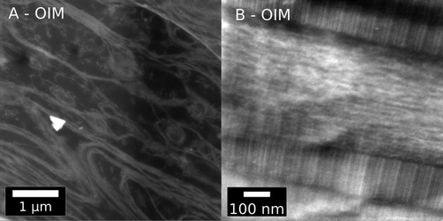
Alternations of collagen and mineral at the molecular level may have a significant impact on the strength and toughness of bone. In this study, scanning transmission electron microscopy (STEM) and electron energy-loss spectroscopy (EELS) were employed to study structural and compositional changes in bone pathology at nanometer spatial resolution. Tail tendon and femoral bone of osteogenesis imperfecta murine (oim, brittle bone disease) and wild type (WT) mice were compared to reveal defects in the architecture and chemistry of the collagen and collagen-mineral composite in the oim tissue at the molecular level. There were marked differences in the substructure and organization of the collagen fibrils in the oim tail tendon; some regions have clear fibril banding and organization, while in other regions fibrils are disorganized. Malformed collagen fibrils were loosely packed, often bent and devoid of banding pattern. In bone, differences were detected in the chemical composition of mineral in oim and WT. While mineral present in WT and oim bone exhibited the major characteristics of apatite, examination in EELS of the fine structure of the carbon K ionization edge revealed a significant variation in the presence of carbonate in different regions of bone. Variations have been also observed in the fine structure and peak intensities of the nitrogen K-edge. These alterations are suggestive of differences in the maturation of collagen nucleation sites or cross-links. Future studies will aim to establish the scale and impact of the modifications observed in oim tissues. The compositional and structural alterations at the molecular level cause deficiencies at larger length scales. Understanding the effect of molecular alterations to pathologic bone is critical to the design of effective therapeutics.
中文翻译:

电子显微镜揭示成骨不全小鼠病理学中纳米尺度的结构和化学变化
胶原蛋白和矿物质在分子水平上的交替可能会对骨骼的强度和韧性产生重大影响。在这项研究中,采用扫描透射电子显微镜(STEM)和电子能量损失谱(EELS)来研究纳米级空间分辨率下骨病理学的结构和组成变化。比较了成骨不全鼠(oim,脆性骨病)和野生型(WT)小鼠的尾腱和股骨,从分子水平揭示了oim组织中胶原蛋白和胶原蛋白矿物质复合物的结构和化学缺陷。oim胶原纤维的亚结构和组织存在明显差异尾腱 一些地区的原纤维条带和组织清晰,而其他地区的原纤维则是无序的。畸形的胶原纤维堆积疏松,经常弯曲且没有条带状图案。在骨骼中,检测到oim和WT中矿物质的化学成分存在差异。WT和OIM中存在矿物质骨骼显示出磷灰石的主要特征,在EELS中对碳K电离边缘的精细结构进行检查后发现,骨骼不同区域中碳酸盐的存在存在显着差异。还已经观察到氮K-边缘的精细结构和峰强度的变化。这些改变暗示胶原成核位点或交联的成熟度的差异。未来的研究旨在确定在卵组织中观察到的修饰的规模和影响。在分子水平上的组成和结构改变导致较大长度尺度上的缺陷。了解分子改变对病理性骨骼的影响对于有效治疗剂的设计至关重要。
更新日期:2016-10-14
中文翻译:

电子显微镜揭示成骨不全小鼠病理学中纳米尺度的结构和化学变化
胶原蛋白和矿物质在分子水平上的交替可能会对骨骼的强度和韧性产生重大影响。在这项研究中,采用扫描透射电子显微镜(STEM)和电子能量损失谱(EELS)来研究纳米级空间分辨率下骨病理学的结构和组成变化。比较了成骨不全鼠(oim,脆性骨病)和野生型(WT)小鼠的尾腱和股骨,从分子水平揭示了oim组织中胶原蛋白和胶原蛋白矿物质复合物的结构和化学缺陷。oim胶原纤维的亚结构和组织存在明显差异尾腱 一些地区的原纤维条带和组织清晰,而其他地区的原纤维则是无序的。畸形的胶原纤维堆积疏松,经常弯曲且没有条带状图案。在骨骼中,检测到oim和WT中矿物质的化学成分存在差异。WT和OIM中存在矿物质骨骼显示出磷灰石的主要特征,在EELS中对碳K电离边缘的精细结构进行检查后发现,骨骼不同区域中碳酸盐的存在存在显着差异。还已经观察到氮K-边缘的精细结构和峰强度的变化。这些改变暗示胶原成核位点或交联的成熟度的差异。未来的研究旨在确定在卵组织中观察到的修饰的规模和影响。在分子水平上的组成和结构改变导致较大长度尺度上的缺陷。了解分子改变对病理性骨骼的影响对于有效治疗剂的设计至关重要。


 京公网安备 11010802027423号