当前位置:
X-MOL 学术
›
Cryst. Growth Des.
›
论文详情
Our official English website, www.x-mol.net, welcomes your
feedback! (Note: you will need to create a separate account there.)
Real-Time Observation of Protein Dense Liquid Cluster Evolution during Nucleation in Protein Crystallization
Crystal Growth & Design ( IF 3.2 ) Pub Date : 2017-02-06 00:00:00 , DOI: 10.1021/acs.cgd.6b01826 Robin Schubert 1 , Arne Meyer 2 , Daniela Baitan 1, 2 , Karsten Dierks 2 , Markus Perbandt 1 , Christian Betzel 1
Crystal Growth & Design ( IF 3.2 ) Pub Date : 2017-02-06 00:00:00 , DOI: 10.1021/acs.cgd.6b01826 Robin Schubert 1 , Arne Meyer 2 , Daniela Baitan 1, 2 , Karsten Dierks 2 , Markus Perbandt 1 , Christian Betzel 1
Affiliation

|
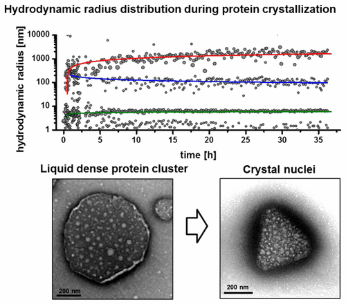
Controlled navigation in the phase diagram of protein crystallization and probing by advanced Dynamic Light Scattering (DLS) technology provided new information and more insight on the early processes during the nucleation process. The observed hydrodynamic radius distribution pattern clearly reveals a two-step mechanism of nucleation and the occurrence of liquid dense protein clusters, which were verified by transmission electron microscopy. The growth kinetics of these protein clusters, forming distinct radii fractions, is analyzed in real time. Further, the data confirmed that critical nuclei show a distinctly different radius distribution than the liquid dense clusters. The data and results provide experimental evidence that during nucleation, a formation of distinct liquid clusters with high protein concentration occur prior to a transition to crystal nuclei by increasing the internal structural order of these clusters, subsequently.
中文翻译:

蛋白质结晶成核过程中蛋白质密集液体簇演化的实时观察
先进的动态光散射(DLS)技术可控制蛋白质结晶和探测相图中的导航,从而提供了新的信息,并提供了有关成核过程中早期过程的更多见解。观察到的流体动力学半径分布规律清楚地揭示了成核和液体致密蛋白簇出现的两步机制,这已通过透射电子显微镜得到了证实。实时分析这些蛋白质簇形成不同半径部分的生长动力学。此外,数据证实临界核显示出与液体密集簇明显不同的半径分布。数据和结果提供了实验证明,在成核过程中,
更新日期:2017-02-06
中文翻译:

蛋白质结晶成核过程中蛋白质密集液体簇演化的实时观察
先进的动态光散射(DLS)技术可控制蛋白质结晶和探测相图中的导航,从而提供了新的信息,并提供了有关成核过程中早期过程的更多见解。观察到的流体动力学半径分布规律清楚地揭示了成核和液体致密蛋白簇出现的两步机制,这已通过透射电子显微镜得到了证实。实时分析这些蛋白质簇形成不同半径部分的生长动力学。此外,数据证实临界核显示出与液体密集簇明显不同的半径分布。数据和结果提供了实验证明,在成核过程中,


 京公网安备 11010802027423号